The anatomy, physiology, and diseases of the membrana pupillaris / by Augustin Prichard.
- Prichard, Augustin, 1818-1898.
- Date:
- 1857
Licence: Public Domain Mark
Credit: The anatomy, physiology, and diseases of the membrana pupillaris / by Augustin Prichard. Source: Wellcome Collection.
Provider: This material has been provided by The Royal College of Surgeons of England. The original may be consulted at The Royal College of Surgeons of England.
3/12
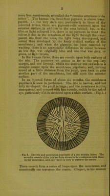
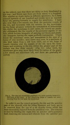
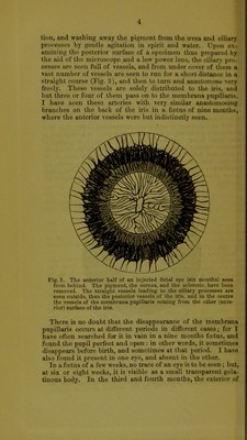
![THE ANATOMY, PHYSIOLOGY, AND DISEASES OF THE MEMBRANA PUPILLARIS. [Bead before the Bath and Bristol Branch, February 19Wi, 1857.] I have ventured to introduce this subject to the notice of the members of the Society, for the purpose of pointing out one or two anatomical errors which, in my opinion, have hitherto ex- isted in the descriptions of the membrana pupillaris and its vessels, and for the sake of bringing forward an opinion which I hold as to the office it serves in foetal life, and also because I wish to narrate a few cases of disease of the eye, which I refer to the continued existence of this organ. The membrana pupillaris, a clear transparent membrane, closing the pupil during foetal life, was first discovered by Wachendorf, in 1738. Afterwards, Albinus, Haller, Zinn, Wrisberg, and Dr. William Hunter, described it about the same time, but no one of them with complete accuracy. It is a simple membrane, continuous with the anterior surface of the iris, quite transparent, and found at every period of foetal life. The centre of the posterior surface is in contact with the an- terior capsule of the lens. Dr. Jacob was the first British author who pointed out that the membrane exists until the ninth month of foetal life; for it had been considered previously not to exist after the seventh—an error originating from the fact that the vessels cease to be permeable at that time; and consequently the membrane is almost invisible, and can only be demonstrated by great care. Tiedemann, Retzius, Arnold, and M. Portal, were also of the opinion that the membrane re- mained till birth. I must call to your remembrance one or two facts respecting the iris, which it is necessary to keep in mind in order to understand the subject, and which, like other anatomical ini notice, are apt to slip away from the recollection, unless there is something to induce us to dwell upon them. In the fully developed and healthy iris, the vessels are posterior, and are covered in behind by the pigment or uvea : the vessels are tortuous and anastomose tolerably freely, the general direction being towards the pupil, where there is a B](https://iiif.wellcomecollection.org/image/b22347288_0005.jp2/full/800%2C/0/default.jpg)