Volume 1
A text-book of human physiology : including histology and microscopical anatomy with special reference to the requirements of practical medicine / by L. Landois ; translated from the seventh German edition with additions by William Stirling.
- Landois, L. (Leonard), 1837-1902. Lehrbuch der Physiologie des Menschen. English
- Date:
- 1891
Licence: Public Domain Mark
Credit: A text-book of human physiology : including histology and microscopical anatomy with special reference to the requirements of practical medicine / by L. Landois ; translated from the seventh German edition with additions by William Stirling. Source: Wellcome Collection.
110/602 page 70
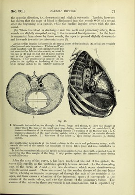
![[The cardiac impulse is synchronous with the systole of the heart, but although this name and apex-beat are frequently used as synonymous terms, it is to be remembered that the impulse may be caused by ditferent parts of the heart being in contact with the chest-wall. The cardiac impulse is usually higher than normal in children, while it is lower during inspira- tion than expiration,] [Methods.—To obtain a curve of the apex-beat or a cardiogram, we may use one or other of the following cardiographs (fig. 43). Fig. 43, A, is the first form used by Marey, and it consists, of an oval wooden capsule ap})lied in an air-tight manner over the apex-beat. The disc, 2h capable of being regulated by the screw, s, presses upon the region of the apex-beat, while t is a tube which may be connected with a lecording tambour (fig. 55). B is an improved form of the instrument, consisting essentially of a tambour, while attached to the membrane is a button, Cardiographs. A, Marey's original form ; B, Marey's improved form ; C, pansphygmo- graph of Brondgeest; D, cardiograph of Burdon-Sandersou; E, that of v. Knoll. p, to be applied over the apex-beat. The movements of the air within the capsule are com- municated by the tube, t, to a recording tambour. Fig. 43, C, is the pansphygmograph of Brondgeest, which consists of a Marey's tambour, in an iron horse-shoe frame, and adjustable by means of a screw, s. Burdon-Sanderson's cardiograph is shown in V. The button, ^j, carried by the spring, e, does not rest upon the caoutchouc membrane, but on an aluminium plate attached to it. The apparatus is adjusted to the chest by three supports. Fig. 43, E, shows a modified instrument on the same principle by Grummach and v.,. Knoll. In all these figures the t indicates the exit-tube communicating with a recording tanibour (fig. 65). D and ^may be used for other purposes, e.g., I'or the pulse, so that they are polygraphs. See also fig. 88.] [For studying the curve of contraction and expansion of the ventricles Roy and Adami used a special myo-cardiograph. Fine hooks were inserted into the ventricular wall, the hooks were attached to threads which hooked over pulleys and were then connected with recording levers. To obtain tracings of the contraction of the papillary nmscles a fine hooked wire was inserted through the auricular wall and hooked over one of the mitral flaps. It slides easily in a collar which is tied to the edges of the opening in the auricular walk To the wire is attached a thread, which, after passing round a pulley, is attached to a recording lever.] Fig 47, A, shows the cardiogram or the impulse-curve of the heart of a healthy man ; B, that of a dog, obtained by means of a sphygmograph. In both, the follow- ing points are to be noticed :—ah, corresponds to the time of the pause and the contraction of the auricles. As the atria contract in the direction of the axis of the heart from the right and above towards the left and below, the apex of the heart moves towards the intercostal space. The two or three smaller elevations are](https://iiif.wellcomecollection.org/image/b20417688_001_0110.jp2/full/800%2C/0/default.jpg)


 No text description is available for this image
 No text description is available for this image
 No text description is available for this image