Volume 1
A text-book of human physiology : including histology and microscopical anatomy with special reference to the requirements of practical medicine / by L. Landois ; translated from the seventh German edition with additions by William Stirling.
- Landois, L. (Leonard), 1837-1902. Lehrbuch der Physiologie des Menschen. English
- Date:
- 1891
Licence: Public Domain Mark
Credit: A text-book of human physiology : including histology and microscopical anatomy with special reference to the requirements of practical medicine / by L. Landois ; translated from the seventh German edition with additions by William Stirling. Source: Wellcome Collection.
112/602 page 72
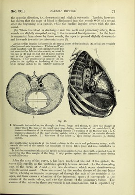
![and upwards {Harvey—'' cor sese erigere), and when hardened during systole presses itself into the intercostal space (fig. 48, II.). (3) The ventricles undergo during systole a slight spiral twisting on their long axis (lateralem inclinationeni —Harvey), so that the apex is brought from behind more for- ward, and thus a greater portion of the left ventricle is turned to the front. This rota- tion is caused by the muscular fibres of the ventricles, which proceed from that part of the fibrous rings between the auricles and ventricles which lies next the anterior thoracic wall. The fibres pass from above obliquely downwards, and to the left, and also run in part upon the posterior surface of the ven- tricles. When they contract in the axis of their direction, they tend to raise the apex, and also to bring more of the.posterior surface Fig. 46. Cardiogram ordog,^shm^^^^^^ ^^^^^ ^^^^^^-^^^ ^^i^j, ^^e anterior points of ^ ^^.^.„f,.— . different observers have referred the thoracic wall It is favoured by the slightly occurrence of the second sound spiral arrangement of the aorta and' pulmonary (closure of the semi-lunar valves). artery. These are the most important causes, but the minor causes are— (4) The reaction impulse or recoil,'' or that movement which the ventricles Fig. 47. Curves from the a]»ex-l:)eat. A, normal curve (man) ; B, from a dog, C, very rapid curve (.log) ; D and E, normal curves (man) registered on a vibrating glass plate Avhere each indentation = 0-01613 sec. In all the curves ab means contraction of the auricles, and be of the ventricles ; d, closure of the aortic, and e, of the pulmonary valves ; ef, diastole of the ventricle. are said to undergo (like an exploded gun or rocket) at the moment when the blood is discharged into the aorta and pulmonary artery, whereby the apex goes in](https://iiif.wellcomecollection.org/image/b20417688_001_0112.jp2/full/800%2C/0/default.jpg)


 No text description is available for this image
 No text description is available for this image
 No text description is available for this image