72 results filtered with: Gummelt, W.
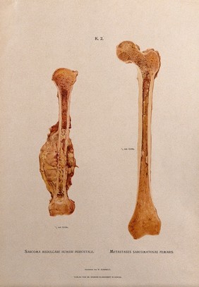
  • Sections through humerus and femur bones, two figures: the left illustration indicating a sarcoma (bone tumour) on the humerus bone, the right showing cancerous (?) metastases in the marrow of the femur. Chromolithograph by W. Gummelt, ca. 1897.
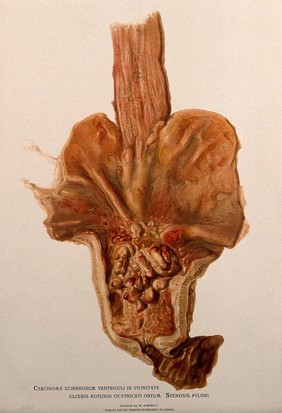
  • A diseased stomach, showing signs of cancer. Chromolithograph by W. Gummelt, ca. 1897.
  • Dissections of pharynxes and larynxes affected by diseases including syphilis. Chromolithograph by W. Gummelt, ca. 1897.
  • Bones and joints deformed by arthritis: five figures. Chromolithograph by W. Gummelt, ca. 1897.
  • Dissection of a diseased intestine. Chromolithograph by W. Gummelt, ca. 1897.
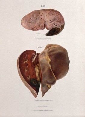
  • Dissections of diseased livers: two figures showing symptoms of amyloidosis hepatis and haemo-siderosis hepatis. Chromolithograph by W. Gummelt, ca. 1897.
  • Hourglass stomach caused by scarring around an ulcer: two figures. Chromolithograph after W. Gummelt, ca. 1897.
  • A diseased oesophagus. Chromolithograph by W. Gummelt, ca. 1897.
  • A diseased liver, showing symptoms of hepatitis interstitialis syphilitica. Chromolithograph by W. Gummelt, ca. 1897.
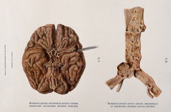
  • Underside of a brain, shown beside a dissection of the abdominal aorta and left iliac artery, both showing symptoms of atherosclerosis and aneurysm. Chromolithograph by W. Gummelt, ca. 1897.
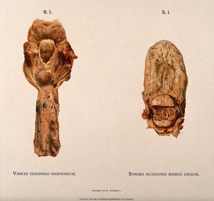
  • Dissections of a diseased oesophagus and tongue: two figures. Chromolithograph by W. Gummelt, ca. 1897.
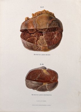
  • Dissections of the spleen affected by infarction of the splenic artery. Chromolithograph by W. Gummelt, ca. 1897.
  • Part of the intestine showing signs of ileitis diphtherica following cholera asiatica. Chromolithograph by W. Gummelt, ca. 1897.
  • Sections through two femur bones: the left illustration indicating the red bone marrow, the right showing the marrow and lymphatics. Chromolithograph by W. Gummelt, ca. 1897.
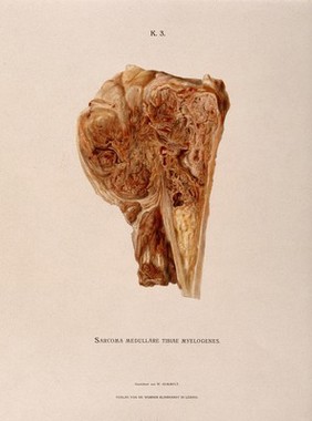
  • Section through part of a tibia bone, showing diseased tissue and a cancerous (?) sarcoma (bone tumour). Chromolithograph by W. Gummelt, ca. 1897.
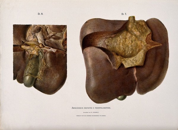
  • A diseased liver with abcesses: two figures showing symptoms of abscessus hepatis e periphlebitide. Chromolithograph by W. Gummelt, ca. 1897.
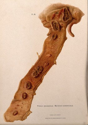
  • Dissection of a diseased intestine, showing signs of typhus. Chromolithograph by W. Gummelt, ca. 1897.
  • Dissections of femoral arteries affected by calcification, with illustrations of subperitoneal phlebectasia and rectal rectal hemorrhoids. Chromolithograph by W. Gummelt, ca. 1897.
  • Dissections of larynxes affected by diseases including cancer: four figures. Chromolithograph by W. Gummelt, ca. 1897.
  • Sections through diseased bones affected by osteochodritis (caused by syphilis), spondylitis and osteomyelitis. Chromolithograph by W. Gummelt, ca. 1897.
  • Dissections of diseased intestines and mesenteries, affected by necrosis, ulcers and tuberculosis: three figures. Chromolithograph by W. Gummelt, ca. 1897.
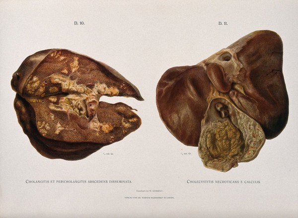
  • Dissections of diseased livers: two figures showing symptoms of cholangitis and cholecystitis. Chromolithograph by W. Gummelt, ca. 1897.
  • A diseased stomach. Chromolithograph by W. Gummelt, ca. 1897.
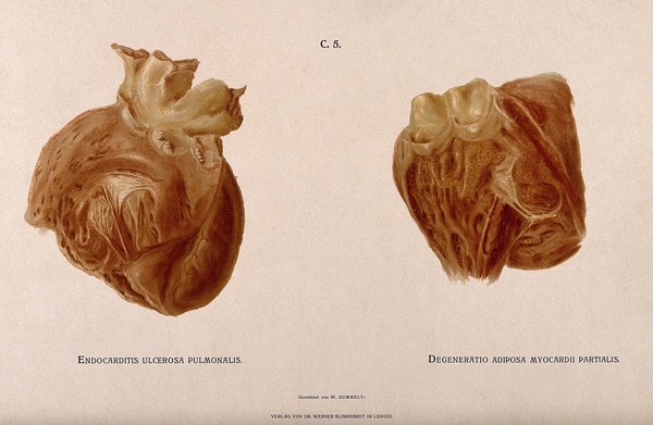
  • Dissections of two diseased hearts. Chromolithograph by W. Gummelt, ca. 1897.
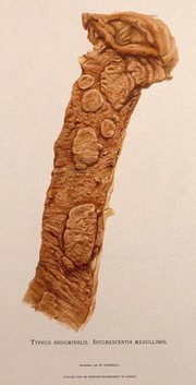
  • Dissection of a diseased intestine, showing signs of typhus. Chromolithograph by W. Gummelt, ca. 1897.
  • A diseased liver, including a section seen under a microscope, showing symptoms of atrophia hepatis acuta. Chromolithograph by W. Gummelt, ca. 1897.
  • Sections through bones affected by Möller-Barlow disease (hypertrophic osteodystrophy). Chromolithograph by W. Gummelt, ca. 1897.
  • A diseased heart affected by a form of endocarditis: two figures. Chromolithograph by W. Gummelt, ca. 1897.
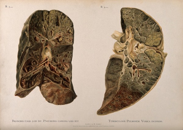
  • Dissections of diseased lungs affected by pneumonia and tuberculosis: two figures. Chromolithograph by W. Gummelt, ca. 1897.
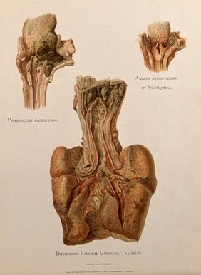
  • Dissections of a diseased pharynx, larynx and tracheae: three figures. Chromolithograph by W. Gummelt, ca. 1897.