Skip to main content
Wellcome Collection homepage
Visit us
What’s on
Stories
Collections
Get involved
About us
Sign in to your library account
Search for anything
Library account
Search for anything
Search
Images search
Search for images
Search
All
All
Catalogue
Catalogue
Images
Images
Events and exhibitions
Events and exhibitions
Stories
Stories
Colours
Licences
Public Domain Mark (59)
Creative Commons CC-BY-NC (13)
Creative Commons CC-BY (5)
In copyright (3)
Dates
From
to
Types/Techniques
Lithography (9)
Engraving and Engravings (7)
Etching (7)
Cartoon (5)
Photograph (5)
Book Illustrations (4)
Albumen prints (3)
Atlases (Scientific) (3)
Bird's-eye view prints (3)
Ephemera (3)
Gelatin silver prints (2)
Portrait prints (2)
Postcard (2)
Allegorical prints (1)
Broadside (1)
Intaglio prints (1)
Panoramic views (1)
Photomechanical prints (1)
Posters (1)
Prints (1)
Subjects
Acquired Immunodeficiency Syndrome (AIDS) (7,565)
HIV Seropositivity (4,295)
Pharmaceutical Preparations (4,031)
Drug Industry (3,975)
London (3,738)
Safe Sex (3,559)
HIV Infections (2,873)
Condoms (2,853)
Botany (2,265)
Human anatomy (1,924)
Plants (1,918)
Hospitals (1,632)
HIV infections - Prevention (1,551)
United Kingdom (1,342)
Costume (1,313)
Death (1,262)
Royal Veterinary College (1,107)
Physicians (1,101)
Paris (1,079)
Charities (1,056)
Dissection (83)
Contributors
Michael Frank, Royal Veterinary College (13)
Charles Heath (9)
Hanhart, Michael, active 1870-1882. (9)
Jean Baptiste François Leveillé (9)
Thomas Pole (4)
James Moores Ball (3)
Scratchley, James, -1849. (3)
Leroux, G. (2)
Stearn Photos (Cambridge) (2)
Alfred Robert Freebairn (1)
Bernard Picart (1)
Bertall (1)
Dent, W., active 1793. (1)
Dorville, Noël, 1874-1938. (1)
Dumont (1)
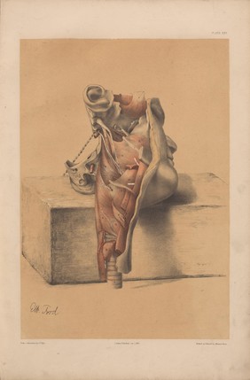
Ford, George H. (George Henry) (1)
George Viner Ellis (1)
Jean-Baptiste Marie Pierre (1)
Luigi Galvani (1)
Mitchell, E., active approximately 1811. (1)
Submit
Active filters:
remove
Dissection
remove
Reset filters
83 results
filtered with: Dissection
Search result sorting
Sort by:
Relevance
Production dates
Sort order:
Ascending
Descending
Submit
Page
1
of 3
Next (page 2)
Close modal window
Page
1
of 3
Next (page 2)


![The London dissector; or, system of dissection, practised in the Hospitals and lecture rooms of the Metropolis ... explained ... comprising a description of the muscles, vessels, nerves, and viscera, of the human body, as they appear on dissection; with directions for their demonstration / [Anon].](https://iiif.wellcomecollection.org/image/L0071161/full/180%2C/0/default.jpg)

![The London dissector; or, system of dissection, practised in the Hospitals and lecture rooms of the Metropolis ... explained ... comprising a description of the muscles, vessels, nerves, and viscera, of the human body, as they appear on dissection; with directions for their demonstration / [Anon].](https://iiif.wellcomecollection.org/image/L0071160/full/180%2C/0/default.jpg)