Skip to main content
Wellcome Collection homepage
Visit us
What’s on
Stories
Collections
Get involved
About us
Sign in to your library account
Search for anything
Library account
Search for anything
Search
Images search
Search for images
Search
All
All
Catalogue
Catalogue
Images
Images
Events and exhibitions
Events and exhibitions
Stories
Stories
Colours
Licences
Creative Commons CC-BY (121)
Creative Commons CC-BY-NC (32)
Creative Commons CC0 (28)
Public Domain Mark (4)
Dates
From
to
Types/Techniques
Books (2)
Broadside (1)
Ephemera (1)
Subjects
Acquired Immunodeficiency Syndrome (AIDS) (7,565)
HIV Seropositivity (4,295)
Pharmaceutical Preparations (4,031)
Drug Industry (3,975)
London (3,738)
Safe Sex (3,559)
HIV Infections (2,873)
Condoms (2,853)
Botany (2,265)
Human anatomy (1,924)
Plants (1,918)
Hospitals (1,632)
HIV infections - Prevention (1,551)
United Kingdom (1,342)
Costume (1,313)
Death (1,262)
Royal Veterinary College (1,107)
Physicians (1,101)
Paris (1,080)
Charities (1,056)
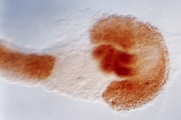
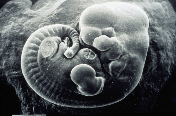
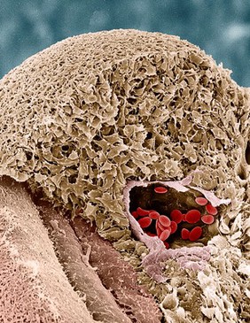
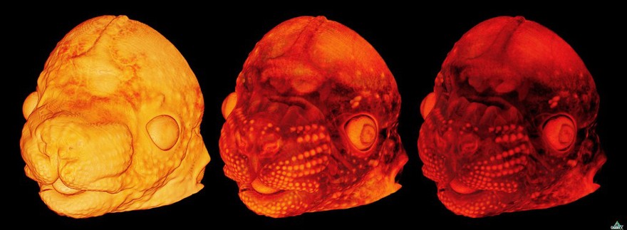
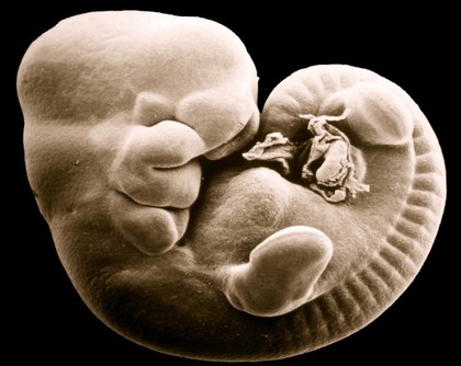
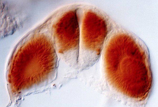
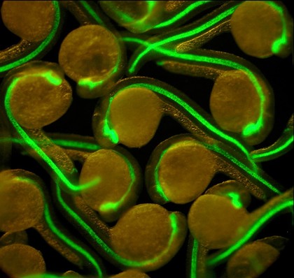
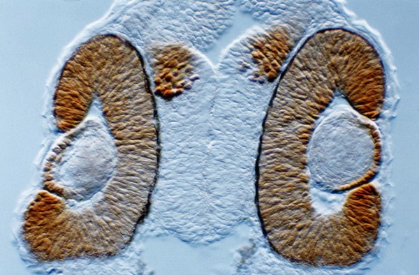
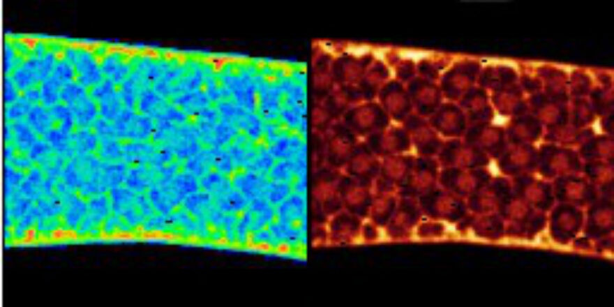
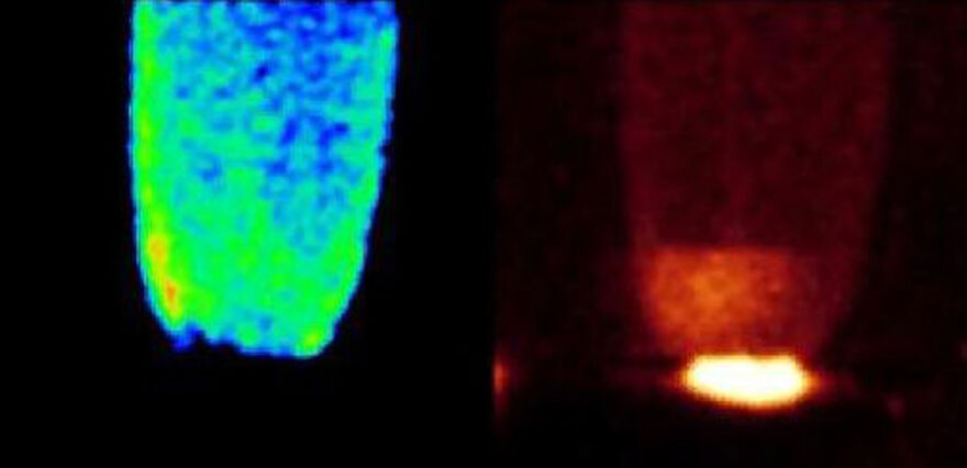
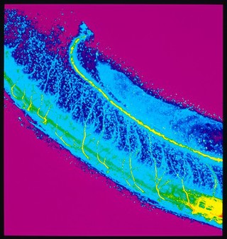
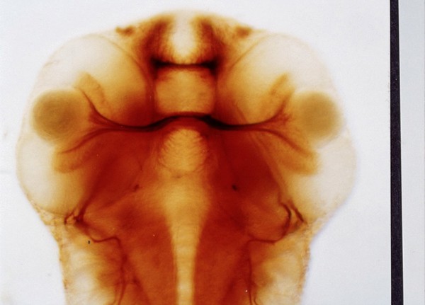
Embryonic Structures (185)
Contributors
Dr Steve Wilson (50)
Huw Parry & Michael Whitaker (22)
Paul Martin (14)
Alan Handyside (12)
Dr Andrea H. Brand (9)
Prof. R. Bellairs (8)
Dr Jonathan Clarke (7)
K. Hardy (7)
Rachel Ashworth/William Hinkes (6)
Dr Mark Carrington,Camb.Univ. (5)
Dr Daniel St. Johnston (4)
Jenny Nichols (4)
Monica Folgueira & Steve Wilson (4)
Anne Weston, Francis Crick Institute (3)
Arwen Wilcock/Univ. Dundee (2)
Hermann Aberle, University of Munster (2)
Kevin Mackenzie, University of Aberdeen (2)
S. Roy & F. Muller (2)
S. Roy & S Higashijima (2)
Uwe Irion & Daniel St Johnston (2)
Submit
Active filters:
remove
Embryonic Structures
remove
Reset filters
185 results
filtered with: Embryonic Structures
Search result sorting
Sort by:
Relevance
Production dates
Sort order:
Ascending
Descending
Submit
Page
1
of 7
Next (page 2)
Close modal window
Page
1
of 7
Next (page 2)