43 results filtered with: Nicolas-Henri Jacob

- Pictures
- Online
Six diagrams illustrating various clubfeet and operations demonstrating how to cure them. Coloured lithograph by J.B.F. Léveillé after N.H. Jacob.
Nicolas-Henri JacobDate: [1831-1854]Reference: 23297i
- Pictures
- Online
Arteries of the thigh. Lithograph by N.H Jacob, 1831/1854.
Nicolas-Henri JacobDate: [1831/1854]Reference: 564162i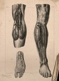
- Pictures
- Online
Arteries of the leg: three figures, one detailing the blood-vesssels of the foot. Lithograph by N.H Jacob, 1831/1854.
Nicolas-Henri JacobDate: [1831/1854]Reference: 564177i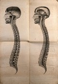
- Pictures
- Online
Cranium and vertebral column: two cross-sections. Lithograph by N.H Jacob, 1831/1854(?).
Nicolas-Henri JacobDate: [1831/1854?]Reference: 564182i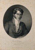
- Pictures
- Online
Charles, Baron Dupin. Lithograph by Langlumé after N.H. Jacob.
Nicolas-Henri JacobDate: 1820Reference: 2675i
- Pictures
- Online
Muscles of the anus: a dissection. Lithograph by N.H Jacob, 1831/1854.
Nicolas-Henri JacobDate: [1831/1854]Reference: 564132i
- Pictures
- Online
Muscles, tendons and bones of the hand: three figures. Lithograph by N.H Jacob, 1831/1854.
Nicolas-Henri JacobDate: [1831/1854]Reference: 564139i
- Pictures
- Online
An operation being performed on the lower abdomen of a male patient. Coloured lithograph by N.H. Jacob after himself.
Nicolas-Henri JacobDate: [1831-1854]Reference: 23284i
- Digital Images
- Online
Plate 17. Surgical instruments for incisions, cauterisations
Nicolas-Henri Jacob
- Digital Images
- Online
Plate 34. Surgical instruments for ligation of arteries.
Nicolas-Henri Jacob
- Digital Images
- Online
Plate H. Surgical technique to correct club foot.
Nicolas-Henri Jacob
- Pictures
- Online
Lymphatic vessels and glands of the human neck and thorax. Lithograph by N.H. Jacob, 1831/1854.
Nicolas-Henri JacobDate: [1831/1854]Reference: 165284i
- Pictures
- Online
Surgical instruments. Coloured lithograph after a drawing by N.H. Jacob.
Nicolas-Henri JacobReference: 493367i
- Pictures
- Online
Dissection of the neck and shoulder, showing the brachial plexus. Lithograph by N.H Jacob, 1831/1854.
Nicolas-Henri JacobDate: [1831/1854]Reference: 564156i
- Pictures
- Online
Muscles and tendons of the foot. Lithograph by N.H Jacob, 1831/1854.
Nicolas-Henri JacobDate: [1831/1854]Reference: 564149i
- Pictures
- Online
Internal anatomy of the neck: two figures, with a black background. Lithograph by N.H Jacob, 1831/1854.
Nicolas-Henri JacobDate: [1831/1854]Reference: 165295i
- Pictures
- Online
An écorché figure, back view, with right arm extended. Lithograph by N.H Jacob, 1831/1854.
Nicolas-Henri JacobDate: [1831/1854]Reference: 564096i
- Pictures
- Online
Arteries of the foot: three figures. Lithograph by N.H Jacob, 1831/1854.
Nicolas-Henri JacobDate: [1831/1854]Reference: 564178i