405 results filtered with: Green
- Digital Images
- Online
3D depth-coloured transparent mouse mammary gland
Felicity Davis, Bethan Lloyd-Lewis and Christine Watson; University of Cambridge
- Digital Images
- Online
Nanographene oxide interacting with bacteria, TEM
Izzat Suffian, Kuo-Ching Mei, Houmam Kafa & Khuloud T. Al-Jamal
- Digital Images
- Online
Adiantum venustum D.Don Adiantaceae (although placed by some in Pteridaceae). Himalayan maidenhair fern. Small evergreen hardy fern. Distribution: Afghanistan-India. It gains its vernacular name from the wiry black stems that resemble hairs. Adiantum comes from the Greek for 'dry' as the leaflets remain permanently dry. The Cherokee used A. pedatum to make their hair shiny. Henry Lyte (1576), writing on A. capillus-veneris, notes that it restores hair, is an antidote to the bites of mad dogs and venomous beasts
Dr Henry Oakeley
- Digital Images
- Online
Lung cancer cells grown in culture, SEM
Anne Weston, Francis Crick Institute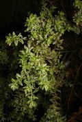
- Digital Images
- Online
Westringia fruticosa 'Variegata'
Dr Henry Oakeley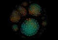
- Digital Images
- Online
Bacterial microbiome mapping, bioartistic experiment
François-Joseph Lapointe, Université de Montréal
- Digital Images
- Online
Mouse Ear Skin
Daniela Malide and Mary Anne Conti, NIH, Bethesda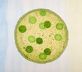
- Digital Images
- Online
Volvox
Odra Noel
- Digital Images
- Online
Rat neurones, SEM
Anne Weston, Francis Crick Institute
- Digital Images
- Online
Situs inversus, illustration
S. Roy
- Digital Images
- Online
Raw onion, illustration
Karen Gustafson
- Digital Images
- Online
Zika virus, illustration
RCSB Protein Data Bank
- Digital Images
- Online
Flesh fly (Sarcophagidae)
Macroscopic Solutions
- Digital Images
- Online
Healthy adult human brain viewed face on, tractography
Henrietta Howells, NatBrainLab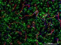
- Digital Images
- Online
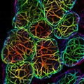
Cellular architecture of normal human skin imaged by whole mount tissue microscopy. Human skin has a rich network of white blood cells (specifically dendritic cells, T cells and macrophages) which form sheaths around blood vessels. This image was taken directly beneath the junction that joins the dermal and epidermal layers of the skin (dermo-epidermal junction). At this level, the capillary network (stained for CD31; red) is visualised against a lawn of autofluorescent dermal papillae (finger-like projections of the dermis; green) scattered with dendritic cells (stained for CD11c; green) and macrophages (stained for LYVE-1; blue). This normal cellular architecture is grossly disrupted in diseased skin (see related images). Scale bar (white) represents 200 micrometres.
Dr. Xiao-nong Wang, Human Dendritic Cell Laboratory, Newcastle University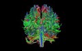
- Digital Images
- Online
Healthy adult human brain viewed from behind, tractography
Henrietta Howells, NatBrainLab
- Digital Images
- Online
Imaginary Herbaria of Dr James Miranda Barry
Alessandra Pirovano
- Digital Images
- Online
Leishmania mexicana parasite in the promastigote stage, SEM
University of Oxford, Richard Wheeler
- Digital Images
- Online
A microCT 3D reconstruction of a 10-day-old chick embryo, as seen from the right hand side. The inner ear is depicted, with the semicircular canals (the body's balance organ) and the cochlea (which converts sound waves into electrical impulses) shown in green. The otic capsule, a cartilaginous structure surrounding the inner ear which develops into part of the sphenoid bone, is shown in blue.
Akshay Kumar, Tom Davies and Nobue Itasaki, University of Bristol
- Digital Images
- Online
Euphorbia characias subsp. wulfenii
Dr Henry Oakeley
- Digital Images
- Online
Bacterial microbiome mapping, bioartistic experiment
François-Joseph Lapointe, Université de Montréal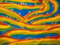
- Digital Images
- Online
ATP sysnthase fields
Odra Noel
- Digital Images
- Online
Cabbage, axial view, MRI
Alexandr Khrapichev, University of Oxford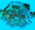
- Digital Images
- Online
Rabbit cerebellum
David Linstead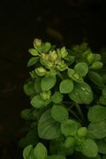
- Digital Images
- Online
Origanum vulgare 'Compactum'
Dr Henry Oakeley