66 results filtered with: Neurone

- Digital Images
- Online
Rat neurones, SEM
Anne Weston, Francis Crick Institute
- Digital Images
- Online
Neurone development, embryoid body
John Grady, Doug Turnbull, Claudia Racca, Newcastle University
- Digital Images
- Online
Neurone development, embryoid body
John Grady, Doug Turnbull, Claudia Racca, Newcastle University
- Digital Images
- Online
Hippocampal neurone, confocal image
Claudius Griesinger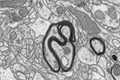
- Digital Images
- Online
Myelinated nerves in a mouse brain, TEM
Mikaela Laine, University of Helsinki
- Digital Images
- Online
Bipolar neurone in the midbrain of an adult zebrafish, LM
Dr Mónica Folgueira
- Digital Images
- Online
Confocal image of neurone + dendrites, BW
Claudius Griesinger
- Digital Images
- Online
Nervous system in a fruit fly larva, serial section TEM
Albert Cardona, HHMI Janelia Research Campus
- Digital Images
- Online
Single neurone in the midbrain of an adult zebrafish, LM
Dr Mónica Folgueira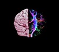
- Digital Images
- Online
Healthy brain, composite of tractography and MRI
Gabriel González-Escamilla
- Digital Images
- Online
Myelinated nerves in a mouse brain, TEM
Mikaela Laine, University of Helsinki
- Digital Images
- Online
Neuroepithelium, the developing brain
Prof. Bill Harris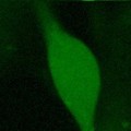
- Digital Images
- Online
Confocal image of neurone, movie.

- Digital Images
- Online
Rat neurones, SEM
Anne Weston, Francis Crick Institute
- Digital Images
- Online
Healthy adult human brain viewed from above, MRI
Dr Flavio Dell'Acqua
- Digital Images
- Online
Healthy adult human brain viewed face on, tractography
Henrietta Howells, NatBrainLab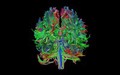
- Digital Images
- Online
Healthy adult human brain viewed from behind, tractography
Henrietta Howells, NatBrainLab
- Digital Images
- Online
Healthy human adult brain viewed from the side, tractography
Dr Flavio Dell'Acqua
- Digital Images
- Online
Symmetric cell division in a live zebrafish embryo
Paula Alexandre, UCL
- Digital Images
- Online
Neuronal synapse, artwork
Stephen Magrath
- Digital Images
- Online
Healthy adult human head and brain viewed from behind, MRI
Dr Flavio Dell'Acqua
- Digital Images
- Online
Neuronal migration is an artwork depicting many very young neurons that have been produced in the neuroepithelium migrating to their appropriate destinations in the brain. This image highlights the future of neuroscience showing different classes of cells colour coded. There is no available technique to do this now, but it is not far off considering the advances that have been made with brainbow mice. The brainbow technique allows for different cell types to be tagged with fluorescent proteins to track their development and connections with other cells.
Prof. Bill Harris
- Digital Images
- Online
Mueller glial cells in the retina
Prof. Bill Harris
- Digital Images
- Online
Cell fates in zebrafish retina, acrylic painting
Prof. Bill Harris
- Digital Images
- Online
Addiction and reward pathways in the brain, artwork
Stephen Magrath