71 results filtered with: T-Lymphocytes
- Books
Monoclonal T cells and their products / edited by Marc Feldmann, Max H. Schreier.
Date: 1982- Books
The handbook of cancer immunology / edited by Harold Waters.
Date: [1978], ©1978- Books
Macrophage regulation of immunity : proceedings of the conference regulatory role of macrophages in immunity, held in Augusta, Michigan, March 12-14, 1979 / edited by Emil R. Unanue, Alan S. Rosenthal.
Date: 1980
- Digital Images
- Online
HIV and antibodies, HIV viral life cycle, illustration
David S. Goodsell, The Scripps Research Institute- Books
Les médicaments antirétroviraux / Rédaction: Jean-Michel Dariosecq.
Date: 2013
- Digital Images
- Online
NK T-cell lymphoma is a highly aggressive cancer of a specific type of immune cell called lymphoid cells, and is associated with the Epstein Barr virus (glandular fever). In later stages of the disease, the lymphoma can spread to the lymph nodes, as in this case.
William R. Geddie
- Digital Images
- Online
Cellular architecture of normal human skin imaged by whole mount tissue microscopy. Human skin has a rich network of white blood cells (specifically dendritic cells, T cells and macrophages) which form sheaths around blood vessels. This image was taken less than 20 micrometres beneath the junction that joins the dermal and epidermal layers of the skin (dermo-epidermal junction). At this level, dendritic cells (stained for CD11c; green) form clusters around and between blood capillary loops (stained for CD31; red). The blind-ended tips of initial lymphatic vessels are just visible (stained for LYVE-1; blue) at this level. This normal cellular architecture is grossly disrupted in diseased skin (see related images). Scale bar (white) represents 200 micrometres.
Dr. Xiao-nong Wang, Human Dendritic Cell Laboratory, Newcastle University
- Digital Images
- Online
Cellular architecture of normal human skin imaged by whole mount tissue microscopy. Human skin has a rich network of white blood cells (specifically dendritic cells, T cells and macrophages) which form sheaths around blood vessels. In this image, T cells (stained for CD3; red) dendritic cells (stained for MHC class II; green) and macrophages (stained for LYVE-1; blue with some cells showing a tinge of green) can be seen. Cell nuclei have been stained with DAPI (grey). This normal cellular architecture is grossly disrupted in diseased skin (see related images). X10 magnification. Scale bar (white) represents 200 micrometres.
Dr. Xiao-nong Wang, Human Dendritic Cell Laboratory, Newcastle University
- Books
- Online
Immunoglobulin idiotypes : proceedings of a symposium held on immunobiological idiotypes and their expression, sponsored by the ICN-UCLA symposia on molecular & cellular biology, held in Salt Lake City, Utah, February 8-15, 1981 / edited by Charles Janeway, Eli E. Sercarz, Hans Wigzell.
Date: 1981- Books
Thymus dependency / edited by A.J.S. Davies and R.L. Carter.
Date: 1973- Books
Idiotypes and lymphocytes / Constantin A. Bona.
Constantin A. BonaDate: 1981- Books
T cell hybridomas / editor, Michael J. Taussig.
Date: [1985], ©1985
- Ephemera
- Online
Uni-Sorb : catalog no. U-01 : ready-to-use nylon wool column for the preparation of enriched T and B cell populations / WAK-Chemie Medical GmbH.
WAK-Chemie Medical (Firm)Date: [1993]
- Digital Images
- Online
DNA sequence of CCR5 Delta 32 gene mutation
Emei Ma, P/C Guy McLoughlin
- Digital Images
- Online
St George and the dragon - T lymphocyte killing a cancer cell
Odra Noel
- Digital Images
- Online
Cellular architecture of normal human skin imaged by whole mount tissue microscopy. Human skin has a rich network of white blood cells (specifically dendritic cells, T cells and macrophages) which form sheaths around blood vessels. In this image, T cells (stained for CD3; red) dendritic cells (stained for MHC class II; green) and macrophages (stained for LYVE-1; blue with some cells showing a tinge of green) can be seen. Cell nuclei have been stained with DAPI (grey). This normal cellular architecture is grossly disrupted in diseased skin (see related images). X20 magnification. Scale bar (white) represents 100 micrometres.
Dr. Xiao-nong Wang, Human Dendritic Cell Laboratory, Newcastle University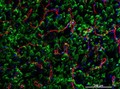
- Digital Images
- Online
Cellular architecture of normal human skin imaged by whole mount tissue microscopy. Human skin has a rich network of white blood cells (specifically dendritic cells, T cells and macrophages) which form sheaths around blood vessels. This image was taken directly beneath the junction that joins the dermal and epidermal layers of the skin (dermo-epidermal junction). At this level, the capillary network (stained for CD31; red) is visualised against a lawn of autofluorescent dermal papillae (finger-like projections of the dermis; green) scattered with dendritic cells (stained for CD11c; green) and macrophages (stained for LYVE-1; blue). This normal cellular architecture is grossly disrupted in diseased skin (see related images). Scale bar (white) represents 200 micrometres.
Dr. Xiao-nong Wang, Human Dendritic Cell Laboratory, Newcastle University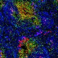
- Digital Images
- Online
Normal spleen showing B cells and T cells
Peter Lane and Fiona McConnell- Books
Monoclonal antibodies and T-cell hybridomas : perspectives and technical advances / editors, Günter J. Hämmerling, Ulrich Hämmerling, and John F. Kearney.
Date: 1981
- Digital Images
- Online
Cellular architecture of normal human skin imaged by whole mount tissue microscopy. Human skin has a rich network of white blood cells (specifically dendritic cells, T cells and macrophages) which form sheaths around blood vessels. In this image, blood vessels (string-like structures stained for CD31; green), lymphatic vessels (ribbon-like structures stained for LYVE-1; blue) and T cells (stained for CD3; red) can be seen. T cells are only found around dermal blood vessels. Macrophages (stained for LYVE-1; blue) are also present. This normal cellular architecture is grossly disrupted in diseased skin (see related images). X10 magnification. Scale bar (white) represents 200 micrometres.
Dr. Xiao-nong Wang, Human Dendritic Cell Laboratory, Newcastle University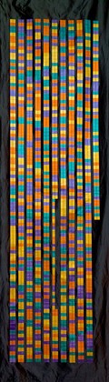
- Digital Images
- Online
DNA sequence of CCR5 Delta 32 gene mutation
Emei Ma, P/C Guy McLoughlin
- Ephemera
- Online
Turn this column upside down : Uni-Sorb ready-to-use nylon wool columns for the preparation fo [sic] enriched T and B cell fractions / WAK-Chemie Medical GmbH.
WAK-Chemie Medical (Firm)Date: [1993]
- Digital Images
- Online
HIV maturation, HIV viral life cycle, illustration
David S. Goodsell, The Scripps Research Institute
- Digital Images
- Online
Complex between T-cell receptor/viral peptid
T.Blundell & N Campillo
- Digital Images
- Online
Dendritic cell interacting with a T cell
Peter Lane and Fiona McConnell