132 results filtered with: Immunology

- Digital Images
- Online
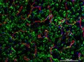
Cellular architecture of normal human skin imaged by whole mount tissue microscopy. Human skin has a rich network of white blood cells (specifically dendritic cells, T cells and macrophages) which form sheaths around blood vessels. In this image, T cells (stained for CD3; red) dendritic cells (stained for MHC class II; green) and macrophages (stained for LYVE-1; blue with some cells showing a tinge of green) can be seen. Cell nuclei have been stained with DAPI (grey). This normal cellular architecture is grossly disrupted in diseased skin (see related images). X20 magnification. Scale bar (white) represents 100 micrometres.
Dr. Xiao-nong Wang, Human Dendritic Cell Laboratory, Newcastle University
- Digital Images
- Online
HeLa cell, immortal human epithelial cancer cell line, SEM
Anne Weston, Francis Crick Institute
- Digital Images
- Online
Lymphocyte with mitochondria
Rob Young
- Digital Images
- Online
Lung cancer cells grown in culture, SEM
Anne Weston, Francis Crick Institute- Books
La passion d'épauler : Albert Calmette, codécouvreur du BCG / Marie-José Hermant, Philippe Scherpereel ; photographies, Sam Bellet.
Hermant, Marie-JoséDate: ©2013
- Digital Images
- Online
'Serum straight from the horse'., inoculation caricature
- Digital Images
- Online
Cellular architecture of normal human skin imaged by whole mount tissue microscopy. Human skin has a rich network of white blood cells (specifically dendritic cells, T cells and macrophages) which form sheaths around blood vessels. This image was taken directly beneath the junction that joins the dermal and epidermal layers of the skin (dermo-epidermal junction). At this level, the capillary network (stained for CD31; red) is visualised against a lawn of autofluorescent dermal papillae (finger-like projections of the dermis; green) scattered with dendritic cells (stained for CD11c; green) and macrophages (stained for LYVE-1; blue). This normal cellular architecture is grossly disrupted in diseased skin (see related images). Scale bar (white) represents 200 micrometres.
Dr. Xiao-nong Wang, Human Dendritic Cell Laboratory, Newcastle University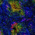
- Digital Images
- Online
Normal spleen showing B cells and T cells
Peter Lane and Fiona McConnell
- Digital Images
- Online
Macrophages infected with candida yeast, LM
Kevin Mackenzie, University of Aberdeen- Books
I.I. Mechnikov i zarozhenie immunologii / T.I. Ulʹi︠a︡nkina.
Ulʹjankina, T. I.Date: 1985- Books
The birth of immunology / Lucien Craps.
Craps, Lucien.Date: [1993], ©1993
- Digital Images
- Online
Human macrophage rupturing after infection with Chlamydia
David Goulding, Wellcome Trust Sanger Institute
- Digital Images
- Online
Cellular architecture of normal human skin imaged by whole mount tissue microscopy. Human skin has a rich network of white blood cells (specifically dendritic cells, T cells and macrophages) which form sheaths around blood vessels. In this image, blood vessels (string-like structures stained for CD31; green), lymphatic vessels (ribbon-like structures stained for LYVE-1; blue) and T cells (stained for CD3; red) can be seen. T cells are only found around dermal blood vessels. Macrophages (stained for LYVE-1; blue) are also present. This normal cellular architecture is grossly disrupted in diseased skin (see related images). X10 magnification. Scale bar (white) represents 200 micrometres.
Dr. Xiao-nong Wang, Human Dendritic Cell Laboratory, Newcastle University- Books
Techniques in clinical immunology / edited by R.A. Thompson.
Date: 1977- Books
Silent signal / Animate Projects ; with scientist Bentley Crudgington.
Animate ProjectsDate: 2016
- Digital Images
- Online
HeLa cells, immortal human epithelial cancer cell line, SEM
Anne Weston, Francis Crick Institute
- Books
- Online
Fighting infection : 4th report / House of Lords, Select Committee on Science and Technology, Session 2002-03.
Great Britain. Parliament. House of Lords. Science and Technology Committee.Date: [2003], ©2003
- Digital Images
- Online
HeLa cell, immortal human epithelial cancer cell line, SEM
Anne Weston, Francis Crick Institute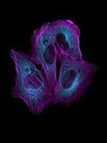
- Digital Images
- Online
HeLa cells, LM
Kevin Mackenzie, University of Aberdeen
- Digital Images
- Online
Dendritic cell interacting with a T cell
Peter Lane and Fiona McConnell
- Books
- Online
Cases of tetanus; and rabies contagiosa, or canine hydrophobia : with remarks, chiefly intended to ascertain the characteristic symptoms of the latter disease in man and certain brutes, and to point out the most effectual means of prevention / by Caleb Hillier Parry.
Parry Caleb Hillier, 1755-1822.Date: 1814- Books
Immunology / Richard A. Goldsby [and others].
Date: [2003], ©2003
- Digital Images
- Online
Dividing HeLa cells, LM
Kevin Mackenzie, University of Aberdeen
- Digital Images
- Online
HeLa cell, immortal human epithelial cancer cell line, SEM
Anne Weston, Francis Crick Institute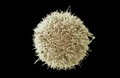
- Digital Images
- Online
HeLa cell, immortal human epithelial cancer cell line, SEM
Anne Weston, Francis Crick Institute