11 results filtered with: Chick

- Digital Images
- Online
Fluorescent cranial nerves
Dr Jonathan Clarke
- Digital Images
- Online
Chick embryo spinal nerves
Dr Jonathan Clarke
- Digital Images
- Online
embryo wound healing SEM
Paul Martin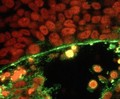
- Digital Images
- Online
Embryonic wound healing
Paul Martin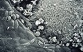
- Digital Images
- Online
embryo wound healing SEM
Paul Martin
- Digital Images
- Online
embryo wound healing SEM
Paul Martin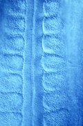
- Digital Images
- Online
Chick embryo somites & SC
Dr Jonathan Clarke
- Digital Images
- Online
A microCT 3D reconstruction of a 10-day-old chick embryo, as seen from the right hand side. The inner ear is depicted, with the semicircular canals (the body's balance organ) and the cochlea (which converts sound waves into electrical impulses) shown in green. The otic capsule, a cartilaginous structure surrounding the inner ear which develops into part of the sphenoid bone, is shown in blue.
Akshay Kumar, Tom Davies and Nobue Itasaki, University of Bristol
- Digital Images
- Online
embryo wound healing SEM
Paul Martin
- Digital Images
- Online
embryonic wound healing
Paul Martin
- Digital Images
- Online
Embryonic chick skeleton
Prof. R. Bellairs