Volume 1
Descriptive catalogue of the preparations in the museum of St. Thomas's Hospital.
- Date:
- 1847-59
Licence: Public Domain Mark
Credit: Descriptive catalogue of the preparations in the museum of St. Thomas's Hospital. Source: Wellcome Collection.
Provider: This material has been provided by Royal College of Physicians, London. The original may be consulted at Royal College of Physicians, London.
69/268 page 49
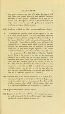
![M.— Nerves. 340. Two nerves from a Frog, one of motion, the other of sensa- tion. The former shows much more distinctly the individual nerve tubules than the latter. 341. Anervewith its neurolemma, enclosing a bundle of nervetubules. 342. Another nerve contained in its neurolemma. 343. Portion of the nervus vagus, showing its nerve tubules filled with coagulated nervous matter. 344. A portion of the lingual, and another of the gustatory nerve. 345. A portion of the ulnar nerve going to a muscle, showing the individual nerve tubules filled with granular matter; also the fact that nerves after the coagulation of their tubular contents, cease to exhibit a double contour. 346. A portion of sympathetic nerve from the mesenteric plexus, showing its great resemblance to fibro-cellular tissue ; it is made up of bundles, of wavy fibres. 347. Sympathetic nerve from a Snake, showing the same kind of structure as in No. 346. N.—Organ of Vision. 348. Iris and choroid coat of a Frog, injected. 349. Choroid coat of a Frog, injected. 350. Another preparation of tlie choroid coat of a Frog, injected. [The three preceding preparations show that tlie vascularity of the choroid membrane is much less than in mammals or in birds.] vol,. I. E](https://iiif.wellcomecollection.org/image/b24758309_0001_0069.jp2/full/800%2C/0/default.jpg)

 No text description is available for this image
 No text description is available for this image
 No text description is available for this image