Volume 1
Descriptive catalogue of the preparations in the museum of St. Thomas's Hospital.
- Date:
- 1847-59
Licence: Public Domain Mark
Credit: Descriptive catalogue of the preparations in the museum of St. Thomas's Hospital. Source: Wellcome Collection.
Provider: This material has been provided by Royal College of Physicians, London. The original may be consulted at Royal College of Physicians, London.
71/268 page 51
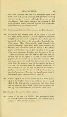
![tion, tliese processes are seen of a triangular figure, with their broad part placed anteriorly, and gradually becoming narrower as they proceed backwards, to be lost on the choroid coat. Each process consists of an extremely rich but single plexus of vessels connected together by a transparent membrane having no definite structure. 361. Membrana 2:)upillaris and ciliary processes of a Kitten, injected. 362. The anterior and posterior layers of the capsule of the lens of a foetal Rabbit, injected. In this preparation, numerous straight vessels can be seen passing from the posterior layer of the capsule of the lens over its margin, into the anterior layer of the capsule. Many of these straight vessels originate in the posterior plexus by a single trunk, which soon divides into two branches, one anastomoses with the vessels of the anterior plexus, and the other seems to pass superficial to the margin of the lens to anastomose, doubtless, with the vessels of the iris. [The preparations, Nos. 353, 354, and 362, show that the anterior layer of the capsule of the lens aud the membrana pupillaris, are in reality one and the same structure. This membrane receives its vessels from two sources, one set pro- ceeds from the posterior layer of the capsule, as seen in No. 362, and the other from the anterior surface of the iris, as seen in Nos. 353 and 354, and whether the membrana pupillaris remain with the iris or with the lens, after the latter has been removed from the eye, depends upon which of these sets of vessels are ruptured in its removal.] .363. Posterior layer of the capsule of the lens of a foetal Kitten, injected, showing tliis plexus to proceed from one large trunk which divides first into two branches, and then these divide each into numerous others wliich radiate from two points, and form, by their subdivisions and anastomoses, a rich plexus. 364. Capsule of the lens of a Kitten, injected. 365. Portion of the lens of a Rabbit. This preparation shows the mode of union of tlie fibres of tlie Lens by their toothed margins, of which its laminiE are made up.](https://iiif.wellcomecollection.org/image/b24758309_0001_0071.jp2/full/800%2C/0/default.jpg)

 No text description is available for this image
 No text description is available for this image
 No text description is available for this image