On the origin and development of the pulps and sacs of the human teeth / by John Goodsir.
- Date:
- [1839?]
Licence: Public Domain Mark
Credit: On the origin and development of the pulps and sacs of the human teeth / by John Goodsir. Source: Wellcome Collection.
Provider: This material has been provided by the Royal College of Physicians of Edinburgh. The original may be consulted at the Royal College of Physicians of Edinburgh.
13/38 page 13
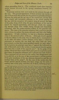
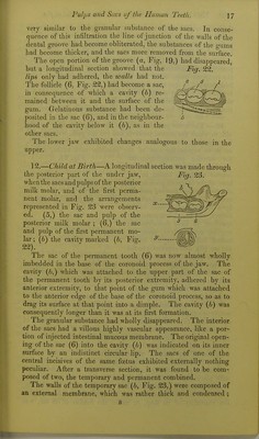
![spongy membrane from the posterior group sliowed, what was not at first observed, that there was at the ])osterior part of the posterior sac another very small one, which by careful examina- tion was seen to be the fundus of the open follicle in the non-ad- herent portion of the dental groove. The adhesion of the lip and walls of the groove had now be- come so strong, that it was impossible to separate them. The only way, therefore, in Avhich its contents could be examined was by transverse sections. When these sections were made between the different sacs, they displayed scarcely any traces of the dental groove ; but when they passed through any ^pifte^-perpendicular toy/- the surface of the gum, and near to the middle of any of the sacs, they exhibited the appearances represented in the marginal sketch, (Fig. 20.) The deciduous tooth pulp, (o,) Mg, 20. which was lately a free papilla; (6,) the section of its sac, which was a follicle when the pulp was a papilla ; (rf,) the line of ad- hesion of part of the walls of the dental groove leading from the shut sac to (c,) the raphe of the groove ; (e,) the section of the non-adherent portion of the groove in the situation of the lunula, which existed behind (/,) the inner laminse of the sac (6,) in its fomier follicular condition. From theconsideration of this section, (Fig. 20,) themodein which the ori- ginal follicle, the non-adherent depression behind the inner lami- nse, and the walls of the dental groove, were connected after full adhesion of all the neighbouring parts, will be easily under- stood. The little cavity (e) adhered by its anterior and inferior extremity to ihe line of adhesion, (d,) so that it and the sac of the milk tooth were both connected to the raphe of the edges of the dental grooves by lines of attachment, which resembled two petioles proceeding from a common footstalk. These lines of at- tachment were not tubular, but resisted all efforts to push a fine probe or bristle through them : they were merely opaque remains of the surfaces of junction contrasting with the semitransparent substance of the gums. Parallel sections through all the sacs ex- hibited similar appearances. When the contents of the sacs were examined, the pulps were found to have acquired the configura- tion of the bodies of the future teeth. The bases by which the molar pulps form.erly adhered to the bottoms of their sacs, and which may be denominated their primary bases, had become al- most divided into three secondary bases, which corresponded with the internal and two external fangs of the future teeth. This di- vision was so far accomplished by the advancement of the internal grey membrane of the sac, under the form of small compressed canals between the base of the pulp and the external spongy mem- brane. These canals, which were three in number, one external](https://iiif.wellcomecollection.org/image/b21955451_0015.jp2/full/800%2C/0/default.jpg)