On the origin and development of the pulps and sacs of the human teeth / by John Goodsir.
- Date:
- [1839?]
Licence: Public Domain Mark
Credit: On the origin and development of the pulps and sacs of the human teeth / by John Goodsir. Source: Wellcome Collection.
Provider: This material has been provided by the Royal College of Physicians of Edinburgh. The original may be consulted at the Royal College of Physicians of Edinburgh.
14/38 page 14
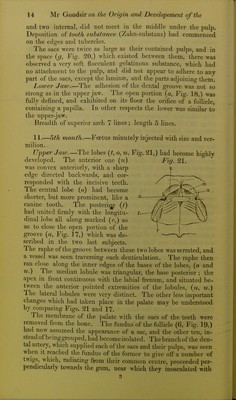
![and two internal, did not meet in the middle under the pulp. Deposition of tooth substance (Zahn-substanz) had commenced on the cdcfes and tubercles. The sacs were twice as large as their contained pulps, and in the space {g. Fig. 20,) which existed between them, there was observed a very soft flocculent gelatinous substance, which had no attachment to the pulp, and did not appear to adhere to any part of the sacs, except the laminaj, and the parts adjoining them. Lower Jaw.—The adhesion of the dental groove was not so strong as in the upper jaw. The open portion (a. Fig. 18,) was fully defined, and exhibited on its floor the orifice of a follicle, containing a papilla. In other respects the lower was similar to the upper-jaw. Breadth of superior arch 7 lines ; length 5 lines. 11.—5th month.—Foetus minutely injected with size and ver- milion. , Upper Jaw.—The lobes {t, o, u, Fig. 21,) had become highly developed. The anterior one {it) Fig. 21. was convex anteriorly, with a sharp edge directed backwards, and cor- responded with the incisive teeth. The central lobe (o) had become shorter, but more prominent, like a canine tooth. The posteri^ (t) had united firmly with the longitu- dinal lobe all along marked (i-,) so as to close the open portion of the groove {a. Fig. ]7,) which was de- scribed in the two last subjects. The raphe of the groove between these two lobes was serrated, and a vessel was seen traversing each denticulation. The raphe then ran close along the inner edges of the bases of the lobes, (o and u.) The median lobule was triangular, the base posterior ; the apex in front continuous with the labial frenum, and situated be- tween the anterior pointed extremities of the lobides, (m, u.) The lateral lobules were very distinct. The other less important changes which had taken place in the palate may be understood by comparing Figs. 21 and 17. The membrane of the palate with the sacs of the teeth were removed from the bone. The fundus of the follicle (6, Fig. 19,) had now assumed the appearance of a sac, and the other ten, in- stead of beinggrouped, had become isolated. The branch of the den- tal artery, which supplied each of the sacs and their pulps, was seen when It reached the fundus of the former to give olFa number of twigs, which, radiating from their common centre, proceeded per- pendicularly towards the gum, near which they inosculated with 3](https://iiif.wellcomecollection.org/image/b21955451_0016.jp2/full/800%2C/0/default.jpg)