The early development of the pericardium, diaphragm, and great veins / by C.B. Lockwood.
- Charles Barrett Lockwood
- Date:
- 1888
Licence: Public Domain Mark
Credit: The early development of the pericardium, diaphragm, and great veins / by C.B. Lockwood. Source: Wellcome Collection.
Provider: This material has been provided by The Royal College of Surgeons of England. The original may be consulted at The Royal College of Surgeons of England.
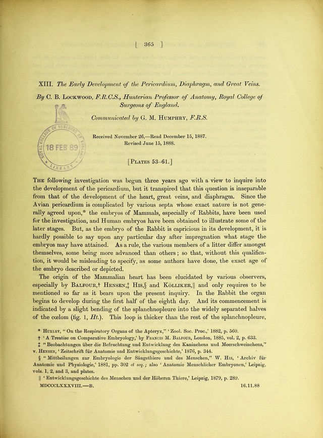
3/40 page 365
![XIII. The Early Development of the Pericardium, Diaphragm, and Great Veins. By C. B. Lockwood, F.R.C.S., Hunterian Professor of Anatomy, Royal College of Surgeons of England. Communicated G. M. Humphry, F.R.S. Received November 26,—Read December 15, 1887. Revised June 15, 1888. [Plates 53-61.] The following investigation was begun three years ago with a view to inquire into the development of the pericardium, but it transpired that this question is inseparable from that of the development of the heart, great veins, and diaphragm. Since the Avian pericardium is complicated by various septa whose exact nature is not gene- rally agreed upon,* * * § the embryos of Mammals, especially of Rabbits, have been used for the investigation, and Human embryos have been obtained to illustrate some of the later stages. But, as the embryo of the Rabbit is capricious in its development, it is hardly possible to say upon any particular day after impregnation what stage the embryos may have attained. As a rule, the various members of a litter differ amongst themselves, some being more advanced than others; so that, without this qualifica- tion, it would be misleading to specify, as some authors have done, the exact age of the embryo described or depicted. The origin of the Mammalian heart has been elucidated by various observers, especially by Balfour,! Hensen,^ His,§ and Kolliker,|| and only requires to be mentioned so far as it bears upon the present inquiry. In the Rabbit the organ begins to develop during the first half of the eighth day. And its commencement is indicated by a slight bending of the splanchnopleure into the widely separated halves of the coelom (fig. 1, Ht.). This loop is thicker than the rest of the splanchnopleure, * Hcjxlet, “ On the Respiratory Organs of the Apteryx,” ‘ Zool. Soc. Proc.,’ 1882, p. 560. t ‘ A Treatise on Comparative Embryology,’ by Francis M. Balfour, London, 1885, vol. 2, p. 633. + “ Beobachtungen iiber die Befruchtung nnd Bntwicklnng des Kaninchens und Meerschweinchens,” V. Hensbn, ‘ Zeitschriffc fiir Anatomie und Bntwicklnngsgeschichte,’ 1876, p. 344. § “ Mittheilungen znr Bmbryologie der Saugethiere und des Menschen,” W. His, ‘ Archiv fill* Anatomie und Physiologie,’ 1881, pp. 302 et seq.; also ‘Anatomie Menschlicher Embryonen,’ Leipzig, vols. 1, 2, and 3, and plates. II ‘ Entwicklnngsgeschichte des Menschen und der Hoheren Thiere,’ Leipzig, 1879, p. 289. MDCCCLXXXVIII.—B. 16.11.88 I .](https://iiif.wellcomecollection.org/image/b22289136_0005.jp2/full/800%2C/0/default.jpg)