On the visual organs in Lamellibranchiata / by Benjamin Sharp.
- Sharp, Benjamin, 1877-1916.
- Date:
- [1883]
Licence: Public Domain Mark
Credit: On the visual organs in Lamellibranchiata / by Benjamin Sharp. Source: Wellcome Collection.
Provider: This material has been provided by UCL Library Services. The original may be consulted at UCL (University College London)
24/28 page 470
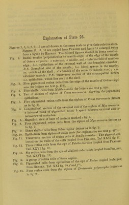
![Explanation of Plate 26. o ft r R 9 16 are all drawn to the same scale to give their relative sizes. Figures 2, 3, 5, 8, 9, lb are au arawu w , fi l5 enlarged twice pjo-nrps 11 12 13 are copied from *raisse anu uguic * o b fg V^'rP hv Hensen The colored figures stained in borax carmine. B?' Branchial ride of the mantle; .... Wood spaces m the mantle; U.S. Bnnomal sio Ietractor muscle (r.«0 ; ..», m 5. Smented retina cells from the siphon of V,nu, zenana ,letters as in fig. 2) eternal end of the siphon of Mya arenaria: ** ]^rlZ7£^^> «™ hereon externa, and in- temal row of tentacles. ' 7. Magnified view of base of tentac^ marked g. 6^ ^ ^ 8. Four pigmented retina cells from the sipnon oi y Fig. Fig. in fig. 2) Fig. 9 _ ^ Jhrfe cells ^t^^t^ see text p. 46,. Fig. 10. Epithelium from siphon of ^ ^^°fcWflPMarirfefl. The pigment col- Transverse section of retina cells °} Taf.XXVIfig.il)- ,ected on the ftomFaatssB, Fig 12 Three retina cells trom tne eye in x« r, .3 «IcS Sm the eye of *»* —«• (copied fromFna.ss, Taf. XXVI fig. 13). Fig. 11. Fig. 16 in fig. 2)](https://iiif.wellcomecollection.org/image/b21642813_0026.jp2/full/800%2C/0/default.jpg)



 No text description is available for this image
 No text description is available for this image
 No text description is available for this image