The Milroy lectures on kala-azar, delivered before the Royal College of Physicians of London. Lecture II, Kala-azar as a disease / by Leonard Rogers.
- Leonard Rogers
- Date:
- 1907
Licence: Public Domain Mark
Credit: The Milroy lectures on kala-azar, delivered before the Royal College of Physicians of London. Lecture II, Kala-azar as a disease / by Leonard Rogers. Source: Wellcome Collection.
5/14 page 490
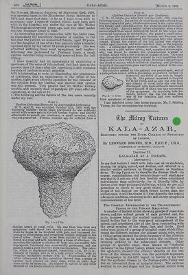
![Term BaitisH MapricaL JovsNaL * Ra the’ Brrrish Mepican JourNAL of December 23rd, 1876, was operated upon in the Manchester Royal Infirmary in 1874, and since that date—as faras I have been able to ascertain—only 2 cases of cystine caleuli have been met with in the hospital, one under the care of Mr. Walter Whitehead about twenty years ago, and the other under the late Professor Jones in 1892. » An interesting point in connexion with the latter case, ‘as illustrating the hereditary character of cystine, is the fact that the patient, an unmarried female, aged 20 years, was the daughter of one of the four patients (also a female) operated upon by my father 24 years previously. She was ‘admitted suffering from renal symptoms, and nephro- lithotomy was performed by Professor Jones, a small cystine calculus being successfully removed from the left kidney. I have recently had an opportunity of examining a specimen of the urine of this patient, and find that at the ‘present time (14 years after the operation) it still contains crystals of cystine in small quantity. titIt is interesting to note, as illustrating the persistence -of cystinuria, that an examination of the urine of her mother, eight years after the operation for the removal of the caleulus from her bladder, also showed that cystine was still present. This patient had no further urinary ‘trouble, and recently died of paralysis (34 years after the -operation), at the age of 67. 4 |The following are the details of the two cases recently under my care: CASE I. Cystine Calculus Removed by Suprapubic Lithotomy. W. S., aged 32, was admitted October 17th, 1906, with the following history: Seven years previously he was suddenly ‘seized with severe pain in the left lumbar region, and shortly afterwards he passed, per urethram, a small calculus, which xvas not preserved. Fifteen months ago he suffered from a \ Line r : sedi AVNG Gira “ys FD siege} DP Aer ped ] tebe ea fi Pea! similar attack of renal colic. He was then free from any ‘symptoms until about two months previous to admission, when he began to be troubled with pain and increased fre- quency in micturition, and occasional sudden stoppage in the dow of urine. Blood had never been noticed in the urine, which was clear and acid, containing atrace ofalbumen. On ‘sounding the bladder a calculus of large size was readily felt. Suprapubic lithotomy was successfully performed, and the patient made a good recovery from the operation. The calcu- lus, which is mushroom-shaped, measuring lin. by lyin., consists of cystine, and is of a pale yellow colour, with an irregular crystalline surface (Fig. 1). Examination of the urine subsequent to the operation showed that crystals of ‘cystine were present on some days, while on other days none could be detected. No history of calculi in any other member of his family could be obtained. |Marcn 2, 1907. CASE Il. Cystine Calculus Passed per Urethram. C. W., 22, single, was admitted October 19th, 1906, with the following history : For about six months she had suffered from frequent attacks of pain, which commenced in the region of the left kidney, and extended across the abdomen and downwards into the thigh. About a month before admission, after a more severe attack than usual, followed by blood in the urine and increased frequency in micturition, she passed a small calcu- lus. After this the attacks of pain became less frequent and less severe, and when admitted into hospital they had almost ceased, though the region of the left kidney was slightly tender upon pressure, and more resistant than on the opposite side. A radiograph gave a negative result. The urine, 1020, acid, was of a pale amber colour, and, examined microscopic- ally, was found to contain the characteristic crystals of cystine, mixed with a few crystals of oxalate oflime. Onsome days the cystine crystals were present in such quantities that they could be seen with the naked eye, forming a deposit upon the cloud of mucus, which settled on standing, and glistening brightly if the specimen glass was held and rotated in the sun- shine or electric light. The calculus passed by the patient was pear-shaped, and of a pale yellow colour, with a glistening crystalline surface; if measured one-third of an inch in length (Fig. 2). The patient remained in hospital for three weeks, and as she was free from any attacks of renal colic she was then discharged, with instructions to return and report herself if there was any recurrence of the symptoms. On inquiring into her family history she was not aware that any of her relatives had ever suffered Che Milroy Wertures & KADLA-AZAR, DELIVERED BEFORE THE RoyaL COLLEGE OF PHYSICIANS or LONDON. By LEONARD ROGERS, M.D., F.R.C.P., I.M.S., PROFESSOR OF PATHOLOGY, CALCUTTA. Lecture II. KALA-AZAR AS A DISEASE, [ABSTRACT. ] In my first lecture I dealt with kala-azar as an epidemic, tracing its origin, spread, and decline, and referred to a similar earlier outbreak in Bengal known as Burdwan fever. To-day I pass on to describe the disease itseli—its course, complications, and terminations—and shall show you that it is not one whit less terrible when considered individually than collectively, for it literally kills by inches after most prolonged sufferings, which we are still powerless to check to any great extent. As the later stages of the disease are much better known than the earlier ones, it will be clearer if I first describe the typical advanced condition, returning to the still rarely recognized commencement of the fever. THE GENERAL APPEARANCES IN THE CHARACTERISTIC STAGES oF THE DISEASE KALA-AZAR, A number of lantern slides showing groups of cases were shown, and the salient points of each pointed out, the main features being the marked contrast between the tumid bellies due to the very great enlargement of the spleen, and sometimes also of the liver, contrasting with the great wasting of the chest, face, and limbs. Brief notes were given of a group of hospital cases which illus- trated the different courses the disease might take, while a village group showed the great frequency of the affection in children, just as in malarial fever, the incidence de- creasing steadily in the subsequent decades. Other groups of sporadic cases, then known as ‘“ malarial cachexia” in the Sylhet Valley, which was not invaded by the epidemic, showed the clinical identity of the two forms of the disease, which was the principal argument of the speaker in his 1897 report in favour of his view that the epidemic disease in the Brahmaputra Valley was but an intensified form of malarial fever. The extreme wasting in the last stages and the dropsical ascitic form](https://iiif.wellcomecollection.org/image/b33455119_0005.jp2/full/800%2C/0/default.jpg)