Fourth report of the Wellcome Tropical Research Laboratories at the Gordon Memorial College, Khartoum. Volume A, Medical / Andrew Balfour.
- Balfour, Andrew
- Date:
- 1911
Licence: Attribution 4.0 International (CC BY 4.0)
Credit: Fourth report of the Wellcome Tropical Research Laboratories at the Gordon Memorial College, Khartoum. Volume A, Medical / Andrew Balfour. Source: Wellcome Collection.
Provider: This material has been provided by London School of Hygiene & Tropical Medicine Library & Archives Service. The original may be consulted at London School of Hygiene & Tropical Medicine Library & Archives Service.
97/442 page 87
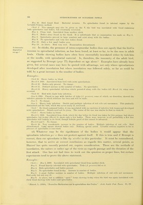
![I fear I cannot answer these questions, but proceed to give illustrations. Unless otherwise stated, all inoculations were made subcutaneously. Bxaiiiplea : Experiments Chicks c and d, clean birds, were inoculated on February 18, 1909, from a case of acute, but not very severe, naturally acquired spirochsetosis in a fowl. On February 20, chick e showed bodies only, and, on the following day, chick d followed suit. The bodies increased in number but at no time was the infection in either bird severe. Chick c, which showed the most bodies, was chloroformed on March 8 and its liver examined by the Levaditi-Manouolian method but no spirochaetes were found. Chick d continued to show bodies for a time but they gradually lessened, and, by March 20, had entirely disappeared from the blood. In neither case was there any marked illness. Chid-, clean bird. Inoculated on May 25 with infected tick tissue (vide infra). On the following day bodies only found in very considerable numbers (there had been no sign of them in the preliminary examination). On May 29, sijirochcetes also were found in the blood. An increase had taken place in the number of bodies. May 30. Increase in the number of spirochajtes. Bodies apjDarently not so numerous as yesterday. May 31. No bodies found even after prolonged search. Spirocheetes fairly numerous. Tangles forming, indicating the crisis. June 1. No spirochsetes. A few bodies. June 3. As above. Jime 6. A few spirochsetes and a few bodies found. Jntin 6. Large numbers of spirochaetes. Tangles again forming. No bodies seen. June 7. As above. June 8. No s])iroch8etes. Bodies present and showing so-called spore forms. June 9 to 13. As above. June 14. Bird died. Nothing special found post mortem. Section of the liver, spleen and lung wore examined by the Lcvaditi-Volpino method, but no sj^irochstes were found. Unfortunately the presence or absence of granules was not noted. This was a most interesting case, and, I am bound to say, suggests a close relationship between the spirochaetes and the inclusions. The alternating presence and absence of the latter is, I think, also suggestive in this direction, as is their increase and decrease shown both in this and other cases {vide infra). (b) In chicks, relapses are common, and the disease therefore approaches mammalian spirochiEtosis in type. Spirochsetes vanish from the peripheral blood, which may then be quite free or may show bodies. Then, the intra-corpuscular bodies, if present, still persisting, the spirochaetes recur. This may happen on several occasions. Examples: Chick X. Clean bird. May 12, 1909. Inoculated with spirochjetal blood from another chick. No bodies present in blood used for inoculation. May 14. Infected. Spirochaetes only. May 15. Slight increase in number of spirochaetes. May 16. Spirochaetes apparently completely absent and remained so till May 18, when they recurred. May 20. Spirochaetes fairly numerous. Chick ill. May 21. Found dead. Spirochastes present in heart's blood. Liver shows a few greenish-yellow spots. Microscopically, there was cloudy swelling and the sjDots were found to be necrotic areas. Chick V. Bird showing some Ijodies in the blood prior to inoculation (vide c and d). Were these true bodies of the after phase ? Apparently so. May 8, 1909. Inoculated with blood from chick showing both sjDirochaetes and bodies. May 9. Both spirochaetes and bodies present. Many of the latter very small. May 10. Enormous number of spirochaetes present. Bodies as above. Bird not very ill. May 11. As above, but tangles numerous to-day. Some of the leucocytes show large, more or less spherical, rose-pink (with Leishman stain) masses in their extra-nuclear i^ortious, the significance of which I am at a loss to understand. Granular white cells, suggesting myelocytes in all but size are present. May 12. As above. May 13. Entire absence of spirochastes. Bodies about the same. May 14. As above. May 15. Recurrence of spirochaetes. Considerable number and tangles. Bodies also increased in number. Multiple infection common. The suggestion is that the spirochfetes are now entering the corpuscles, but this was not actually observed. In the fresh blood, motility of what, at the time, were thought to be genuine bodies was observed. In stained tilms, spore forms found. One corpuscle seen with a gap in it and, hard by, 15 small chromatin granules observed which may well be spores. (Plate III., fig. 3.) A few, though each is distinct, are imited, the majority are free. Possibly, some of the very tiny bodies in the corpuscles are spores which had re-entered the'red cells. This would account for changes in the size of the bodies which may undergo schizogonic development from the merozoite stage.](https://iiif.wellcomecollection.org/image/b21363201_0101.jp2/full/800%2C/0/default.jpg)


 No text description is available for this image
 No text description is available for this image
 No text description is available for this image