The treatment of fractures / Charles Locke Scudder ... Assisted by Frederic J. Cotton, M.D.
- Charles Locke Scudder
- Date:
- 1901
Licence: Public Domain Mark
Credit: The treatment of fractures / Charles Locke Scudder ... Assisted by Frederic J. Cotton, M.D. Source: Wellcome Collection.
Provider: This material has been provided by the Augustus C. Long Health Sciences Library at Columbia University and Columbia University Libraries/Information Services, through the Medical Heritage Library. The original may be consulted at the the Augustus C. Long Health Sciences Library at Columbia University and Columbia University.
178/468 (page 174)
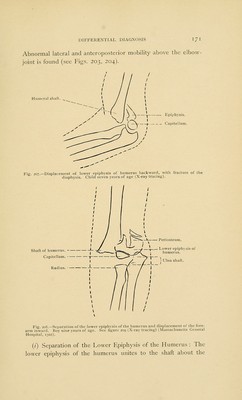
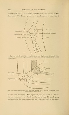
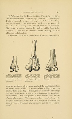
![fracture above the condyles. The diagnosis is made upon the following points : The age of the individual ; the history of the accident; the existence of abnormal mobility at a very low level on the humeral shaft; anteroposterior mobility very Olecranon fossa. Interna] portion of epiphysis. Ulna. — -— __ Humeral epiphysis and —. bits from the diaphy- sis. — - Capitellnm. — - Radial epiphysis. — Radius. Fig. 213.—Separation of the lower epiphysis of the humerus, after union. Anteropos- terior view. This figure illustrates the fact that the epiphysis does not include the condyles of the humerus (X-ray tracing). Z3fcf+- United humeral epiphysis. Capitellnm. 1 1 Radial epiphysis. Fig. 214.—Separation of the lower humeral epiphysis, after union. Lateral view. Extension normal. Flexion to a right angle (X-ray tracing) (Massachusetts General Hospital, 1556). marked, lateral mobility being less marked ; muffled crepitus (this term is very suggestive, and is used by Poland). The breadth of the lower end of the humeral fragment is broader than in the case of a fracture (see Figs. 207 to 214 inclusive).](https://iiif.wellcomecollection.org/image/b21207690_0178.jp2/full/800%2C/0/default.jpg)