Morris's human anatomy : a complete systematic treatise by English and American authors / ed. by C.M. Jackson eleven hundred and eighty two illustrations, three hundred and fifty eight printed in colors.
- Sir Henry Morris, 1st Baronet
- Date:
- [1914]
Licence: Public Domain Mark
Credit: Morris's human anatomy : a complete systematic treatise by English and American authors / ed. by C.M. Jackson eleven hundred and eighty two illustrations, three hundred and fifty eight printed in colors. Source: Wellcome Collection.
Provider: This material has been provided by the Augustus C. Long Health Sciences Library at Columbia University and Columbia University Libraries/Information Services, through the Medical Heritage Library. The original may be consulted at the the Augustus C. Long Health Sciences Library at Columbia University and Columbia University.
101/1564 page 81
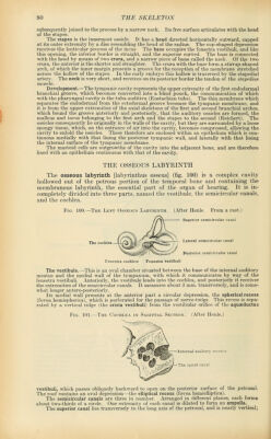
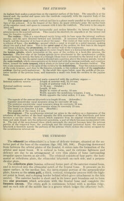
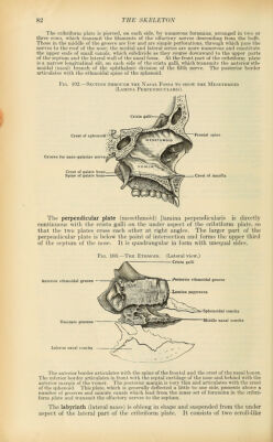
![its highest limb makes a projection on the superior surface of the bone. The ampulla is at the lateral end; the medial end opens into the vestibule conjointly with the superior limb of the posterior canal. The posterior canal is nearly vertical and lies in a plane nearly parallel to the posterior sur- face of the petrosal. It is the longest of the three; its upper extremity joins the medial hmb of the superior canal, and opens in common with it into the vestibule. The lower is the ampul- lated end. The lateral canal is placed horizontally and arches laterally; its lateral limb forms a prominence in the mastoid antrum. This canal is the shortest; its ampulla is at the lateral end near the fenestra vestibuli. The cochlea.—This is a cone-shaped cavity lying with its base upon the internal auditory meatus, and the apex directed forward and laterally. It measures about five millimetres in length, and the diameter of its base is about the same. The centre of this cavity is occupied by a column of bone—the modiolus—around which a canal is wound in a spiral manner, making about two and a half turns. This is the spiral canal of the cochlea; its first turn is the largest and forms a bulging, the promontory, on the medial wall of the tympanum. Projecting into the canal throughout its entire length there is a horizontal,shelf-like lamella, the lamina spiralis, which terminates at the apex of the cochlea in a hook-like process, the hamulus. The free edge of the lamina spiralis gives attachment to the membranous cochlea, a canal having in section the form of a triangle whose base is attached to the lateral wall of the spiral canal. By this the spiral canal is divided into a portion above the lamina spiralis, termed the scaia vestibuli, which communicates at its lower end with the osseous vestibule, and a portion below, termed the scala tympani, which abuts at its lower end upon the fenestra cochlea. The two scalae communicate at the apex of the cochlea by the helicotrema. Near the commencement of the scala tympani, and close to the fenestra rotunda, is the cochlear orifice of the canaliculus cochlese (ductus perilymphaticus). In the adult this opens below, near the middle of the pos- terior border of the petrous bone, and transmits a small vein from the cochlea to the jugular fossa. Measurements of the principal parts connected with the auditory organs:— Internal auditory meatus Length of anterior wall, 13-14 mm. Length of posterior waU, 6.7 mm. External auditory meatus 14^16 mm. (Gruber.) Tympanum Length, 13 mm. Height in centre of cavity, 15 mm. Width opposite the membrana tympani, 2 mm. Width opposite the tubal orifice, 3-4 mm. (Von Troltsch.) The capsule of the osseous labyrinth is in length 22 mm. (Schwalbe.) Superior semicircular canal measures along its convexity 20 mm. The posterior semicircular canal measures along its convexity 22 mm. The lateral semicircular canal measures along its convexity 15 mm. The canal is in diameter 1.5 mm. (Huschke.) The ampulla of the canal, 2.5 mm. Development.—The membranous internal ear arises in the embryo as a depression of the ectoderm of the surface of the head opposite the fifth neuromere of the hind-brain and later becomes a sac-like cavity, the otocyst, which separates from its original ectodermal connec- tions and sinks deeply into the subjacent mesoderm, a part of which becomes incorporated with it. The rest of the mesodermal tissue which surrounds the otocyst becomes later the petrous portion of the temporal bone, the perilymph and the internal periosteal layer; the osseous labyrinth is therefore merely the portions of the petrous which enclose the cavity occupied by the membranous internal ear. THE ETHMOID The ethmoid [os ethmoidale] is a bone of delicate texture, situated at the an- terior part of the base of the cranium (figs. 102, 103, 104). Projecting downward from between the orbital plates of the frontal, it enters into the formation of the orbital and nasal fossae. It is cubical in form, and its extreme lightness and delicacy are due to an arrangement of very thin plates of bone surrounding irregular spaces known as air-cells. The ethmoid consists of four parts: the hori- zontal or cribriform plate, the ethmoidal labyrinth on each side, and a perpen- dicular plate. The cribriform plate [lamina cribrosa] forms part of the anterior cranial fossa, and is received into the ethmoidal notch of the frontal bone. It presents on its upper surface, in the median line, the intra-cranial portion of the perpendicular plate, known as the crista galli, a thick, vertical, triangular process with the high- est point in front, and a sloping border behind which gives attachment to the falx cerebri. The anterior border is short and in its lower part broadens out to form two alar processes which articulate with the frontal bone and complete the foramen caecum. The ci'ista galli is continuous behind with a median ridge, and on each side of the middle line is a groove which lodges the olfactory bulb.](https://iiif.wellcomecollection.org/image/b21212600_0101.jp2/full/800%2C/0/default.jpg)
 No text description is available for this image
 No text description is available for this image
 No text description is available for this image