Morris's human anatomy : a complete systematic treatise by English and American authors / ed. by C.M. Jackson eleven hundred and eighty two illustrations, three hundred and fifty eight printed in colors.
- Sir Henry Morris, 1st Baronet
- Date:
- [1914]
Licence: Public Domain Mark
Credit: Morris's human anatomy : a complete systematic treatise by English and American authors / ed. by C.M. Jackson eleven hundred and eighty two illustrations, three hundred and fifty eight printed in colors. Source: Wellcome Collection.
Provider: This material has been provided by the Augustus C. Long Health Sciences Library at Columbia University and Columbia University Libraries/Information Services, through the Medical Heritage Library. The original may be consulted at the the Augustus C. Long Health Sciences Library at Columbia University and Columbia University.
105/1564 page 85
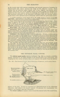
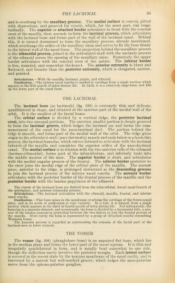
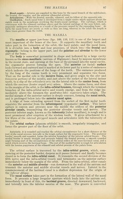
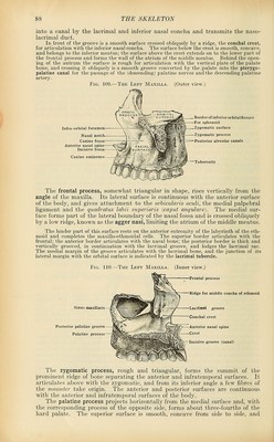
![and is overhung by the maxillary process. The medial surface is convex, pitted with depressions, and grooved for vessels, which, for the most part, run longi- tudinally. The superior or attached border articulates in front with the conchal crest of the maxilla, then ascends to form the lacrimal process, which articulates with the lacrimal bone and forms part of the wall of the lacrimal canal. Behind this, it is turned downward to form the maxillary process, already mentioned, which overhangs the orifice of the maxillary sinus and serves to fix the bone firmly to the lateral wall of the nasal fossa. The projection behind the maxillary process is the ethmoidal process, joined in the articulated skull with the uncinate process of the ethmoid across the opening of the maxillary sinus. Posteriorly the upper border articulates with the conchal crest of the palate. The inferior border is free, rounded, and somewhat thickened. The anterior extremity is blunt and flattened, and broader than the posterior extremity, which is elongated, narrow, Ossification.—The inferior nasal conotia is ossified in cartilage from a single nucleus which appears in the fifth month of intra-uterine life. At birth it is a relatively large bone and filla THE LACRIMAL The lacrimal bone [os lacrimale] (fig. 105) is extremely thin and delicate, quadrilateral in shape, and situated at the anterior part of the medial wall of the orbit. It is the smallest of the facial bones. The orbital surface is divided by a vertical ridge, the posterior lacrimal crest, into two unequal portions. The anterior, smaller portion is deeply grooved to form the lacrimal groove, which lodges the lacrimal sac and forms the com- mencement of the canal for the naso-lacrimal duct. The portion behind the ridge is smooth, and forms part of the medial wall of the orbit. The ridge gives origin to the orbicularis oculi (pars lacrimalis) muscle and ends below in a hook-like process, the lacrimal hamulus, which curves forward to articulate with the lacrimal tubercle of the maxilla and completes the superior orifice of the naso-lacrimal (lacrimo-ethmoidal), forms part of the infundibulum, and inferiorly looks into the middle meatus of the nose. The superior border is short, and articulates with the medial angular process of the frontal. The inferior border posterior to the crest joins the medial edge of the orbital plate of the maxilla. The narrow piece, anterior to the ridge, is prolonged downward as the descending process to join the lacrimal process of the inferior nasal concha. The anterior border articulates with the posterior border of the frontal process of the maxilla and the posterior border with the lamina papyracea of the ethmoid. The vessels of the lacrimal bone are derived from the infra-orbital, dorsal nasal branch of the ophthalmic, and anterior ethmoidal arteries. Articulations.—The lacrimal articulates with the ethmoid, maxilla, frontal, and inferior nasal concha. Ossification.—This bone arises in the membrane overlying the cartilage of the fronto-nasal plate, and in its mode of ossification is very variable. As a rule, it is formed from a single nucleus which appears in the third or fourth month of intra-uterine life. Not infrequently, the hamulus is a separate element, and occasionally the bone is divided by a horizontal cleft, a pro- cess of the lamina papyracea projecting between the two halves to join the frontal process of the maxilla. More rarely the bone is represented by a group of detached ossicles resembling Wormian bones. The hamular process is regarded as representing the remains of the facial part of the lacrimal seen in lower animals. THE VOMER The vomer (fig. 106) (ploughshare bone) is an unpaired flat bone, which lies in the median plane and forms the lower part of the nasal septum. It is thin and irregularly quadrilateral in form, and is usually bent somewhat to one side, though the deflection rarely involves the posterior margin. Each lateral surface is covered in the recent state by the mucous membrane of the nasal cavity, and is traversed by a narrow but well-marked groove, which lodges the naso-palatine nerve from the spheno-palatine ganglion.](https://iiif.wellcomecollection.org/image/b21212600_0105.jp2/full/800%2C/0/default.jpg)
 No text description is available for this image
 No text description is available for this image
 No text description is available for this image