Morris's human anatomy : a complete systematic treatise by English and American authors / ed. by C.M. Jackson eleven hundred and eighty two illustrations, three hundred and fifty eight printed in colors.
- Sir Henry Morris, 1st Baronet
- Date:
- [1914]
Licence: Public Domain Mark
Credit: Morris's human anatomy : a complete systematic treatise by English and American authors / ed. by C.M. Jackson eleven hundred and eighty two illustrations, three hundred and fifty eight printed in colors. Source: Wellcome Collection.
Provider: This material has been provided by the Augustus C. Long Health Sciences Library at Columbia University and Columbia University Libraries/Information Services, through the Medical Heritage Library. The original may be consulted at the the Augustus C. Long Health Sciences Library at Columbia University and Columbia University.
107/1564 page 87
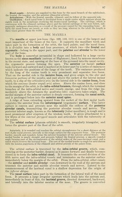
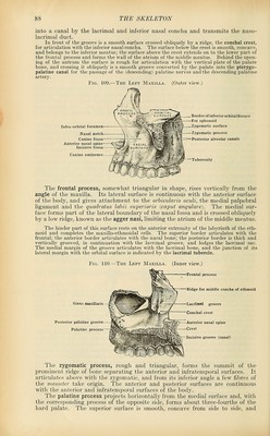
![Blood-supply.—Arteries are supplied to this bone by the nasal branch of the ophthalmic, the frontal, the angular, and the anterior ethmoidal arteries. Articulations.—With the frontal, maxilla, ethmoid, and its fellow of the opposite side. Ossification.—Each nasal bone is developed from a single centre which appears about the eighth week in the membrane overlying the fronto-nasal cartilage. The cartilage, which is continuous with the ethmoid cartilage above and the lateral cartilage of the nose below, sub- sequently undergoes absorption as a result of the pressure caused by the expanding bone. A.t birth the nasal bones are nearly as wide as they are long, whereas in the adult the length is three times greater than the width. THE MAXILLA The maxilla or upper jaw-bone (figs. 109, 110, 111) is one of the largest and most important of the bones of the face. It supports the maxillary teeth and takes part in the formation of the orbit, the hard palate, and the nasal fossa. It is divisible into a body and four processes, of which two—the frontal and zygomatic—belong to the upper part, and the palatine and alveolar to the lower part of the bone. The body is somewhat pyramidal in shape and hollowed by a large cavity known as the sinus maxillaris (antrum of Highmore), lined by mucous membrane in the recent state, and opening at the base of the pyramid into the nasal cavity, the zygomatic process forming the apex. The anterior (or facial) surface looks forward and outward and is marked at its lower part by a series of eminences which indicate the positions of the fangs of the teeth. The eminence produced by the fang of the canine tooth is very prominent and separates two fossa. That on the medial side is the incisive fossa, and gives origin to the alar and transverse portions of the nasalis, and just above the socket of the lateral incisor tooth, to a slip of the orbicularis oris; on the lateral side is the canine fossa, from which the caninus {levator anguli oris) arises. Above the canine fossa, and close to the margin of the orbit, is the infra-orbital foramen, through which the terminal branches of the infra-orbital nerve and vessels emerge, and from the ridge im- mediately above the foramen the quadratus labii superioris takes origin. The medial margin of the anterior surface is deeply concave, forming the nasal notch, and is prolonged below into the anterior nasal spine. A ridge of bone extending upward from the socket of the first molar tooth separates the anterior from the infratemporal (zygomatic) surface. This latter surface is convex and presents near the middle the orifices of the posterior alveolar canals, transmitting the posterior alveolar vessels and nerves. The posterior inferior angle, known as the tuberosity [tuber maxillare], is rough and is most prominent after eruption of the wisdom tooth. It gives attachment to a few fibres of the internal pterygoid muscle and articulates with the tuberosity of the palate. The orbital surface [planum orbitale] is smooth, irregularly triangular, and forms the greater part of the floor of the orbit. Anteriorly, it is rounded and reaches the orbital circumference for a short distance at the root of^the nasal process; lateraUy is the rough surface for the zygomatic bone. The posterior border, smooth and rounded, forms the inferior boundary of the inferior orbital fissure. The medial border is nearly straight and presents behind the frontal process, a smooth rounded angle forming part of the circumference of the orbital orifice of the naso-lacrimal canal, and a notch which receives the lacrimal bone. The rest of the medial border is rough for articulation with the lamina papyracea of the ethmoid and orbital process of the palate bone. The orbital surface is traversed by the infra-orbital groove, which, com- mencing at the posterior border, deepens as it passes forward and finally becomes closed in to form the infra-orbital canal. It transmits the second division of the fifth nerve and the infra-orbital vessels and terminates on the anterior surface immediately below the margin of the orbit. From the infra-orbital, other canals —the anterior and middle alveolar—run downward in the wall of the antrum and transmit the anterior and middle alveolar vessels and nerves. Lateral to the commencement of the lacrimal canal is a shallow depression for the origin of the inferior oblique. The nasal surface takes part in the formation of the lateral wall of the nasal fossa. It presents a large irregular aperture which leads into the antrum and, immediately in front of this, the lacrimal groove, directed downward, backward, and laterally into the inferior meatus of the nose. The groove is converted (](https://iiif.wellcomecollection.org/image/b21212600_0107.jp2/full/800%2C/0/default.jpg)
 No text description is available for this image
 No text description is available for this image
 No text description is available for this image