Morris's human anatomy : a complete systematic treatise by English and American authors / ed. by C.M. Jackson eleven hundred and eighty two illustrations, three hundred and fifty eight printed in colors.
- Sir Henry Morris, 1st Baronet
- Date:
- [1914]
Licence: Public Domain Mark
Credit: Morris's human anatomy : a complete systematic treatise by English and American authors / ed. by C.M. Jackson eleven hundred and eighty two illustrations, three hundred and fifty eight printed in colors. Source: Wellcome Collection.
Provider: This material has been provided by the Augustus C. Long Health Sciences Library at Columbia University and Columbia University Libraries/Information Services, through the Medical Heritage Library. The original may be consulted at the the Augustus C. Long Health Sciences Library at Columbia University and Columbia University.
117/1564 page 97
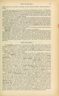
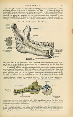
![The condylar process consists of the condyle [capitulum mandibulse] and the narrowed portion by which it is supported, the neck. The condyle is oval in shape, with its long axis transverse to the upper border of the ramus, but oblique with regard to the median axis of the sicull, so that the lateral extremity, which presents the condylar tubercle for the temporo-mandibular ligament of the temporo-mandibular articulation, is a little more forward than the medial ex- tremity. The convex surface of the condyle is covered with cartilage in the recent Fig. 121.—The Mandible. (Medial view.) Lin^ula Mandibular foramen Spheno-mandib- ular ligament Superior constnctor Mylo-hyoid groove Internal pterygoid Stylo-mandibular ligament ^ > tii {—Groove for sub- lingual gland Genio-glossus Genio-hyoid Digastric Mylo-hyoid Groove for submaxillary gland Mylo-hyoid line state, and rests in the mandibular fossa; the neck is flattened from before back- ward, and presents, in front, a depression [fovea pterygoidea] for the insertion of the external pterygoid muscle. The coronoid process, flattened and triangular, is continued upward from the anterior part of the ramus. The lateral surface is smooth and gives insertion to the temporal and masseter muscles; the medial surface is marked by a ridge which descends from the tip and becomes continuous with the posterior part of the mylo-hyoid line. On the medial surface, as well as on the tip of the coronoid Fig 122—Mandiblb showing Relations op Meckel's Cartilage in Human Fcetus op 8 CM. Cbown-Rump Length. (After KoUmann, Handatlas der Entwickelungsgeschichte.) Groove for teeth Meckel's cartilage AnniUus tym- panicus process, the temporal muscle is inserted. The mandibular notch, the deep semi- lunar excavation separating the coronoid from the condylar process, is crossed by the masseteric nerve and vessels. Blood-supply.—Compared with other bones, the superficial parts of the mandible are not so freely supplied with blood. The chief artery is the inferior alveolar which runs in the man- dibular canal, and hence, as the bone is exposed to injury and sometimes actually laid bare in its alveolar portion, it often necroses, especially if the artery is involved at the same time. 7](https://iiif.wellcomecollection.org/image/b21212600_0117.jp2/full/800%2C/0/default.jpg)
 No text description is available for this image
 No text description is available for this image
 No text description is available for this image