Morris's human anatomy : a complete systematic treatise by English and American authors / ed. by C.M. Jackson eleven hundred and eighty two illustrations, three hundred and fifty eight printed in colors.
- Sir Henry Morris, 1st Baronet
- Date:
- [1914]
Licence: Public Domain Mark
Credit: Morris's human anatomy : a complete systematic treatise by English and American authors / ed. by C.M. Jackson eleven hundred and eighty two illustrations, three hundred and fifty eight printed in colors. Source: Wellcome Collection.
Provider: This material has been provided by the Augustus C. Long Health Sciences Library at Columbia University and Columbia University Libraries/Information Services, through the Medical Heritage Library. The original may be consulted at the the Augustus C. Long Health Sciences Library at Columbia University and Columbia University.
129/1564 page 109
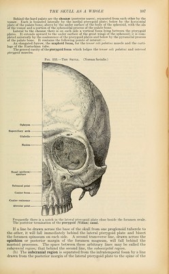
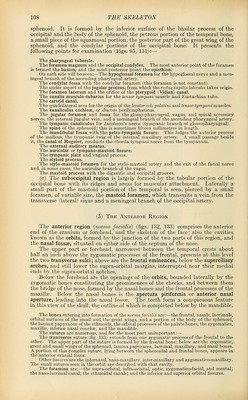
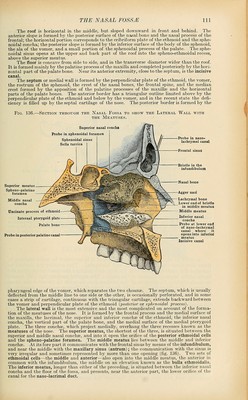
![The following points may also be noticed:— The glabella, a smooth space between the converging superciliary arches. The ophryon, the most anterior point of the metopic suture. The nasion, the middle of the naso-frontal sutui'e. The subnasal point, the middle of the inferior border of the pyriform aperture at the base of the nasal spine. The alveolar point, the centre of the anterior margin of the upper alveolar arch. THE ORBITS The orbits [orbitse] (fig. 134) are two cavities of pyramidal shape, with their bases directed forward and laterally and their apices backward and medially; their medial walls are nearly parallel, but their lateral walls diverge so as to be nearly at right angles to each other. Each cavity forms a socket for the eyeball and the muscles, nerves, and vessels associated with it. Seven bones enter into formation of its walls, viz., the frontal, zygomatic, sphenoid, ethmoid, lacrimal, palate, and maxilla; but as three of these—the frontal, sphenoid, and ethmoid—are single median bones which form parts of each cavity, there are only eleven bones represented in the two orbits. Each orbit presents for examination four walls, a circumference or base, and an apex. The superior wall or roof, vaulted and smooth, is formed mainly by the orbital plate of the frontal and is completed posteriorly by the small wing of the sphenoid. At the lateral angle it presents the lacrimal fossa for the lacrimal gland, and at the medial angle a depression or a spine for the puOey of the superior oblique muscle. Fig. 134.—The Medial Wall of the Orbit. Frontal process of maxilla / V T.arrimfll- /— Lacrimal canal Orifice of antrum Inferior nasal concha Palate b( Anterior nasal spine Anterior ethmoid canal Posterior ethmoid canal Optic foramen Lamina papyracea of ethmoid \ r^Spheno-palatine foramen ?^—^Pterygoid canal, leading into the ^^^ pterygo-palatine fossa Sphenoid External pterygoid plate The inferior wall or floor is directed upward and laterally and is not so large as the roof. It is formed by the orbital plate of the maxilla, the orbital process of the zygomatic, and the orbital process of the palate bone. At its medial angle it presents the naso-laorimal canal, and near this, a depression for the origin of the inferior oblique muscle. It is marked near the middle by a furrow for the infra-orbital artery and the second division of the fifth nerve, terminating anteriorly in the infra-orbital canal, through which the nerve and artery emerge on the face. Near the commencement of the canal a narrow passage, the anterior alveolar canal, runs for- ward and downward in the anterior wall of the antrum, transmitting nerves and vessels to the incisor and canine teeth. The lateral wall, directed forward and medially, is formed by the orbital surface of the great wing of the sphenoid, and the zygomatic. Between it and the roof, near the apex, is the superior orbital (sphenoidal) fissure, by means of which the third, fourth, ophthalmic division of the fifth, and sixth nerves enter the orbit from the cranial cavity; it also transmits some filaments from the cavernous plexus of the sympathetic, the orbital branch of the middle men- ingeal artery, recurrent branches of the lacrimal artery, and an ophthalmic vein. The lower margin of the fissure presents near the middle a small tubercle, from which the inferior head of the lateral rectus muscle arises. Between the lateral wall and the floor, near the apex, is the inferior orbital (spheno-maxiUary) fissure, tlu-ough which the second division of the fifth and the infra-orbital vessels pass from the pterygo-palatine fossa to enter the infra-orbital groove. At the anterior margin of the fissure the sphenoid occasionally articulates with the maxilla, but](https://iiif.wellcomecollection.org/image/b21212600_0129.jp2/full/800%2C/0/default.jpg)
 No text description is available for this image
 No text description is available for this image
 No text description is available for this image