Morris's human anatomy : a complete systematic treatise by English and American authors / ed. by C.M. Jackson eleven hundred and eighty two illustrations, three hundred and fifty eight printed in colors.
- Sir Henry Morris, 1st Baronet
- Date:
- [1914], [©1914]
Licence: Public Domain Mark
Credit: Morris's human anatomy : a complete systematic treatise by English and American authors / ed. by C.M. Jackson eleven hundred and eighty two illustrations, three hundred and fifty eight printed in colors. Source: Wellcome Collection.
Provider: This material has been provided by the Augustus C. Long Health Sciences Library at Columbia University and Columbia University Libraries/Information Services, through the Medical Heritage Library. The original may be consulted at the the Augustus C. Long Health Sciences Library at Columbia University and Columbia University.
50/1564 (page 30)
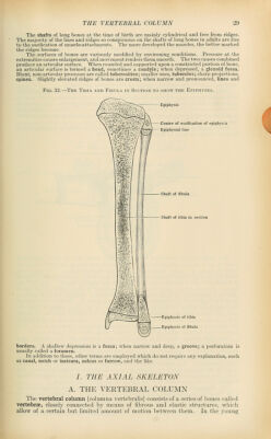
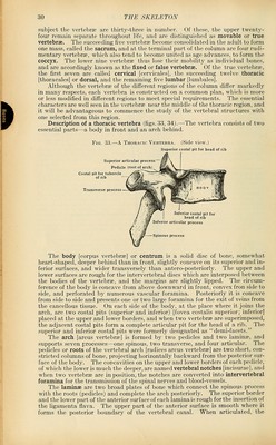
![subject the vertebrse are thirty-three in number. Of these, the upper twenty- four remain separate throughout life, and are distinguished as movable or true vertebrae. The succeeding five vertebrae become consolidated in the adult to form one mass, called the sacrum, and at the terminal part of the column are four rudi- mentary vertebrae, which also tend to become united as age advances, to form the coccyx. The lower nine vertebrae thus lose their mobihty as individual bones, and are accordingly known as the fixed or false vertebrae. Of the true vertebrae, the first seven are called cervical [cervicales], the succeeding twelve thoracic [thoracales] or dorsal, and the remaining five lumbar [lumbales]. Although the vertebrae of the different regions of the coliunn differ markedly in many respects, each vertebra is constructed on a common plan, which is more or less modified in different regions to meet special requirements. The essential characters are well seen in the vertebrae near the middle of the thoracic region, and it will be advantageous to commence the study of the vertebral structures with one selected from this region. Description of a thoracic vertebra (figs. 33, 34).—The vertebra consists of two essential parts—a body in front and an arch behind. Fia. 33.—-A Thoracic Vertebra. (Side view.) Superior costal pit for head of i Superior articular process Pedicle (root of Transverse process Inferior costal pit for head of rib Inferior articular process -Spinous process The body [corpus vertebrae] or centrum is a soUd disc of bone, somewhat heart-shaped, deeper behind than in front, slightly concave on its superior and in- ferior surfaces, and wider transversely than antero-posteriorly. The upper and lower surfaces are rough'for the intervertebral discs which are interposed between the bodies of the vertebrae, and the margins are slightly lipped. The circum- ference of the body is concave from above downward in front, convex fron side to side, and perforated by numerous vascular foramina. Posteriorly it is concave from side to side and presents one or two large foramina for the exit of veins from the cancellous tissue. On each side of the body, at the place where it joins the arch, are two costal pits (superior and inferior) [fovea costalis superior; inferior] placed at the upper and lower borders, and when two vertebrae are superimposed, the adjacent costal pits form a complete articular pit for the head of a rib. The superior and inferior costal pits were formerly designated as demi-facets. The arch [arcus vertebrae] is formed by two pedicles and two laminae, and supports seven processes—one spinous, two transverse, and four articular. The pedicles or roots of the vertebral arch [radices arcus vertebrae] are two short, con- stricted columns of bone, projecting horizontally backward from the posterior sur- face of the body. The concavities on the upper and lower borders of each pedicle, of which the lower is much the deeper, are named vertebral notches [incisurae], and when two vertebrae are in position, the notches are converted into intervertebral foramina for the transmission of the spinal nerves and blood-vessels. The laminae are two broad plates of bone which connect the spinous process with the roots (pedicles) and complete the arch posteriorly. The superior border and the lower part of the anterior surface of each lamina is rough for the insertion of the ligamenta flava. The upper part of the anterior surface is smooth where it forms the posterior boundary of the vertebral canal. When articulated, the](https://iiif.wellcomecollection.org/image/b21212600_0050.jp2/full/800%2C/0/default.jpg)