Morris's human anatomy : a complete systematic treatise by English and American authors / ed. by C.M. Jackson eleven hundred and eighty two illustrations, three hundred and fifty eight printed in colors.
- Sir Henry Morris, 1st Baronet
- Date:
- [1914]
Licence: Public Domain Mark
Credit: Morris's human anatomy : a complete systematic treatise by English and American authors / ed. by C.M. Jackson eleven hundred and eighty two illustrations, three hundred and fifty eight printed in colors. Source: Wellcome Collection.
Provider: This material has been provided by the Augustus C. Long Health Sciences Library at Columbia University and Columbia University Libraries/Information Services, through the Medical Heritage Library. The original may be consulted at the the Augustus C. Long Health Sciences Library at Columbia University and Columbia University.
79/1564 page 59
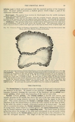
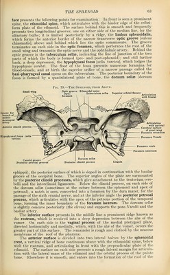
![inferior angle is thick and articulates with the mastoid portion of the temporal bone. Its inner surface presents a shallow groove which lodges a part of the transverse (lateral) sinus. Blood-supply.—The parietal bone receives its blood-supply from the middle meningeal, occipital, and supra-orbital arteries. Articulations.—The parietal articulates with the occipital, frontal, sphenoid, temporal, its fellow of the opposite side, and the epipterio bone when present. Occasionally the temporal and epipteric bones exclude the parietal from articulation with the great wing of the sphenoid. Ossification.—The parietal ossifies from a single nucleus which appears in the outer layer of the membranous wall of the skull about the seventh week. The ossification radiates in such a way as to leave a cleft at the upper part of the bone in front of the occipital angle, the Fig. 74.—Unusual Form op Pabietal Exhibiting a Horizontal Suture Separating the Bone into Two Pif.ces, Upper and Lower. ^f^^^^^^^sy^m cleft of the two side forming a lozenge-shaped space across the sagittal suture known as the sag- ittal fontanelle. This is usually closed about the fifth month of intra-uterine life, but traces may sometimes be recognised at the time of birth, and the parietal foramina are to be regarded as remains of the cleft. According to Dr. A. W. W. Lea, a well-developed sagittal fontanelle is present in 4.4 per cent, of infants at birth. In such cases it closes within the first two months of life, but at times it may remain open for at least eight months after birth and possibly longer. Rarely the parietal bone is composed of two pieces (fig. 74), one above the other, and separated by an antero-posterior suture (sub-sagittal suture), more or less parallel with the sagittal suture. In such cases the parietal is ossified from two centres of ossification. THE FRONTAL The frontal bone [os frontale] closes the cranium in front and is situated above the skeleton of the face. It consists of two portions—a frontal {vertical) portion [squama frontalis], forming the convexity of the forehead, and an orbital {hori- zontal) portion, which enters into formation of the roof of each orbit. Frontal {vertical) portion.—The frontal surface is smooth and convex, and usually presents in the middle line above the root of the nose some traces of the suture which in young subjects traverses the bone from the upper to the lower part. This suture, known as the frontal or metopic suture, indicates the line of junction of the two lateral halves of which the bone consists at the time of birth; in the adult the suture is usually obliterated except at its lowest part. On each side is a rounded elevation, the frontal eminence [tuber frontale], very prominent in young bones, below which is a shallow groove, the sulcus transversus, separat- ing the frontal eminence from the superciliary arch. The latter forms an arched pi^ojection above the margin of the orbit and corresponds to an air-cavity within the bone known as the frontal sinus; it gives attachment to the orbicularis oculi and the corrugator muscles. The ridges of the two sides converge toward the '](https://iiif.wellcomecollection.org/image/b21212600_0079.jp2/full/800%2C/0/default.jpg)
 No text description is available for this image
 No text description is available for this image
 No text description is available for this image