Morris's human anatomy : a complete systematic treatise by English and American authors / ed. by C.M. Jackson eleven hundred and eighty two illustrations, three hundred and fifty eight printed in colors.
- Sir Henry Morris, 1st Baronet
- Date:
- [1914]
Licence: Public Domain Mark
Credit: Morris's human anatomy : a complete systematic treatise by English and American authors / ed. by C.M. Jackson eleven hundred and eighty two illustrations, three hundred and fifty eight printed in colors. Source: Wellcome Collection.
Provider: This material has been provided by the Augustus C. Long Health Sciences Library at Columbia University and Columbia University Libraries/Information Services, through the Medical Heritage Library. The original may be consulted at the the Augustus C. Long Health Sciences Library at Columbia University and Columbia University.
88/1564 page 68
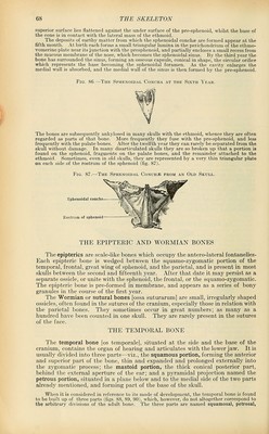
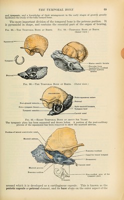
![superior surface lies flattened against the under surface of the pre-sphenoid, whilst the base of the cone is in contact with the lateral mass of the ethmoid. The deposits of earthy matter from which the sphenoidal conchae are formed appear at the fifth month. At birth each forms a small triangular lamina in the peiichondrium of the ethmo- vomerine plate near its junction with the presphenoid, and partially encloses a small recess from the mucous membrane of the nose, which becomes the sphenoidal sinus. By the third year the bone has surrounded the sinus, forming an osseous capsule, conical in shape, the circular orifice which represents the base becoming the sphenoidal foramen. As the cavity enlarges the medial wall is absorbed, and the medial wall of the sinus is then formed by the pre-sphenoid. Fig. 86.—The Sphenoidal Concha at the Sixth Year. The bones are subsequently ankylosed in many skulls with the ethmoid, whence they are often regarded as parts of that bone. More frequently they fuse with the pre-sphenoid, and less frequently with the palate bones. After the twelfth year they can rarely be separated from the skull without damage. In many disarticulated skulls they are so broken up that a portion is found on the sphenoid, fragments on the palate bones, and the remainder attached to the ethmoid. Sometimes, even in old skulls, they are represented by a very thin triangular plate on each side of the rostrum of the sphenoid (fig. 87). Fig. 87.—The Sphenoidal Conch.s: from an Old Skull. Sphenoidal concha Rostrum of sphenoid THE EPIPTERIC AND WORMIAN BONES The epipterics are scale-like bones which occupy the antero-lateral fontanelles- Each epipteric bone is wedged between the squamo-zygomatic portion of the temporal, frontal, great wing of sphenoid, and the parietal, and is present in most skulls between the second and fifteenth year. After that date it may persist as a separate ossicle, or unite with the sphenoid, the frontal, or the squamo-zygomatic. The epipteric bone is pre-formed in membrane, and appears as a series of bony granules in the course of the first year. The Wormian or sutural bones [ossa suturarum] are small, irregularly shaped ossicles, often found in the sutures of the cranium, especially those in relation with the parietal bones. They sometimes occur in great numbers; as many as a hundred have been counted in one skull. They are rarely present in the sutures of the face. THE TEMPORAL BONE The temporal bone [os temporale], situated at the side and the base of the cranium, contains the organ of hearing and articulates with the lower jaw. It is usually divided into three parts—viz., the squamous portion, forming the anterior and superior part of the bone, thin and expanded and prolonged externally into the zygomatic process; the mastoid portion, the thick conical posterior part, behind the external aperture of the ear; and a pyramidal projection named the petrous portion, situated in a plane below and to the medial side of the two parts already mentioned, and forming part of the base of the skull. When it is considered in reference to its mode of development, the temporal bone is found to be built up of three parts (figs. 88, 89, 90), which, however, do not altogether correspond to the arbitrary divisions of the adult bone. The three parts are named squamosal, petrosal,](https://iiif.wellcomecollection.org/image/b21212600_0088.jp2/full/800%2C/0/default.jpg)
 No text description is available for this image
 No text description is available for this image
 No text description is available for this image