Morris's human anatomy : a complete systematic treatise by English and American authors / ed. by C.M. Jackson eleven hundred and eighty two illustrations, three hundred and fifty eight printed in colors.
- Sir Henry Morris, 1st Baronet
- Date:
- [1914]
Licence: Public Domain Mark
Credit: Morris's human anatomy : a complete systematic treatise by English and American authors / ed. by C.M. Jackson eleven hundred and eighty two illustrations, three hundred and fifty eight printed in colors. Source: Wellcome Collection.
Provider: This material has been provided by the Augustus C. Long Health Sciences Library at Columbia University and Columbia University Libraries/Information Services, through the Medical Heritage Library. The original may be consulted at the the Augustus C. Long Health Sciences Library at Columbia University and Columbia University.
90/1564 page 70
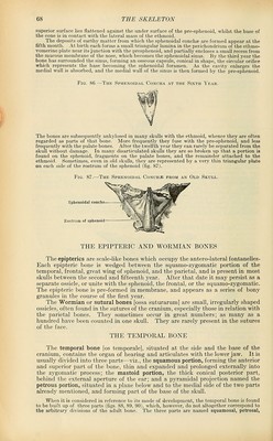
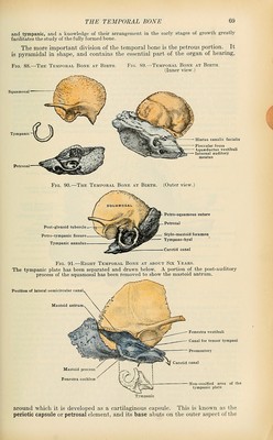
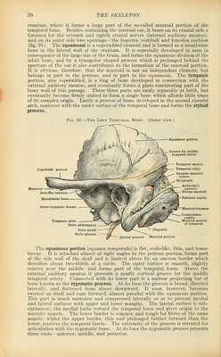
![cranium, where it forms a large part of the so-called mastoid portion of the temporal bone. Besides containing the internal ear, it bears on its cranial side a foramen for the seventh and eighth cranial nerves (internal auditory meatus), and on its outer side two openings—the fenestra vestibuli and fenestra cochleae (fig. 91). The squamosal is a superadded element and is formed as a membrane bone in the lateral wall of the cranium. It is especially developed in man in consequence of the large size of the brain, and forms the squamous division of the adult bone, and by a triangular shaped process which is prolonged behind the aperture of the ear it also contributes to the formation of the mastoid portion. It is obvious, therefore, that the mastoid is not an independent element, but belongs in part to the petrous, and in part to the squamous. The tympanic portion, also superadded, is a ring of bone developed in connection with the external auditory meatus, and eventually forms a plate constituting part of the bony wall of this passage. These three parts are easily separable at birth, but eventually become firmly united to form a single bone which affords little trace of its complex origin. Lastly a process of bone, developed in the second visceral arch, coalesces with the under surface of the temporal bone and forms the styloid process. Fig. 92.—The Left Temporal Bone. (Outer view.) Zygomatic process Tympanic plati Stylo-pharyngeus Stylo-hyoid Stylo-glossus Styloid process Mastoid process The squamous portion [squama temporalis] is flat, scale-like, thin, and trans- lucent. It is attached almost at right angles to the petrous portion, forms part of the side wall of the skull and is limited above by an uneven border which describes about two-thirds of a circle. The outer surface is smooth, slightly convex near the middle, and forms part of the temporal fossa. Above the external auditory meatus it presents a nearly vertical groove for the middle temporal artery. Connected with its lower part is a narrow projecting bar of bone known as the zygomatic process. At its base the process is broad, directed lateralljr, and flattened from above downward. It soon, however, becomes twisted on itself and runs forward, almost parallel with the squamous portion. This part is much narrower and compressed laterally so as to present medial and lateral surfaces with upper and lower margins. The lateral surface is sub- cutaneous; the medial looks toward the temporal fossa and gives origin to the masseter muscle. The lower border is concave and rough for fibres of the same muscle, whilst the upper border, thin and prolonged further forward than the lower, receives the temporal fascia. The extremity of the process is serrated for articulation with the zygomatic bone. At its base the zygomatic process presents three roots—anterior, middle, and posterior.](https://iiif.wellcomecollection.org/image/b21212600_0090.jp2/full/800%2C/0/default.jpg)
 No text description is available for this image
 No text description is available for this image
 No text description is available for this image