A manual of operative surgery / By Lewis A. Stimson.
- Lewis Atterbury Stimson
- Date:
- 1885
Licence: Public Domain Mark
Credit: A manual of operative surgery / By Lewis A. Stimson. Source: Wellcome Collection.
Provider: This material has been provided by the Augustus C. Long Health Sciences Library at Columbia University and Columbia University Libraries/Information Services, through the Medical Heritage Library. The original may be consulted at the the Augustus C. Long Health Sciences Library at Columbia University and Columbia University.
69/536 page 63
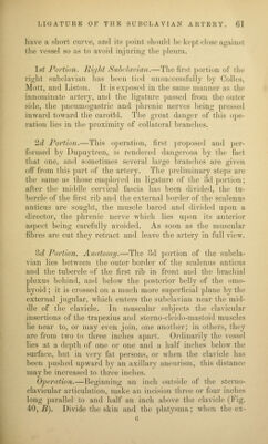
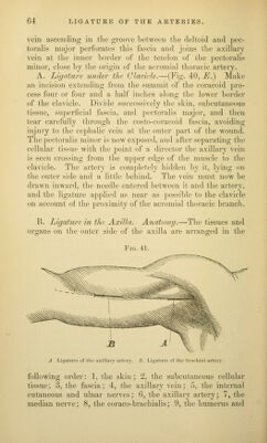
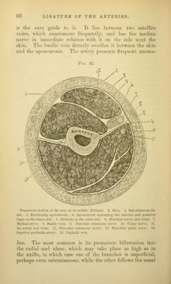
![tlio carotid tu])cTC'U', and seek tlic arlcry Ik'Iow it, scjtaratiii;!; tlic cellular tissue with a director. i*ass the needle between the artery and vein. LIGATURE QF TJIE VERTEBRAL ARTERY. Anatomy.—The vertebral artery passes from the first portion of the subclavian upward and backward to the transverse process of the sixth cervical vertebra. It is ac- companied by a vein which lies in front, and is covered by the deep cervical fascia. The guide to it is the carotid tubercle. Operation.—The first incision is the same as for ligature of the inferior thyroid (Fig. 40, D). The anterior edge of the sterno-cleido-mastoid is exposed and drawn outward. The middle fiiscia is divided, and the carotid and jugular drawn inward. The gap between the longus colli and the scalenus anticus is then felt for about half an inch below the carotid tubercle, the deep fascia covering it torn through, the muscles separated, the vertebral vein pushed aside, and the artery exposed. Chassaignac prefers an incision along the posterior border of the mastoid muscle, and reaches the carotid tubercle by drawing the muscle and vessels inward. If the muscle is very broad some of its clavicular fibres must be divided. LIGATURE OF THE AXILLARY ARTERY. Anatomy.—T\n2 axillary extends from the middle of the clavicle to the lower edge of the tendon of the teres major. The axillary vein lies on the inner side and in front of it. and the brachial nerves invest its lower portion closely. It can be tied below the clavicle in the clavi-pectoral triangle formed by the clavicle, inner border of the i)ectoralis minor, and the thorax, or in the axilla. The strong fascia which unites the coracoid ])rocess and clavicle, and forms the sus- pensory ligament of the axilla, the costo-coracoid fiiscia, sends a prolongation about the upper portion of the axillary vein which keeps its walls from sinking in; the cephalic](https://iiif.wellcomecollection.org/image/b21206533_0069.jp2/full/800%2C/0/default.jpg)
 No text description is available for this image
 No text description is available for this image
 No text description is available for this image