Round celled sarcoma of the anterior mediastinum : extensive metastases, including the brain, both choroid coats, oculo-motor and optic nerves, and external ocular muscles / by Arthur V. Meigs and G. E. de Schweinitz.
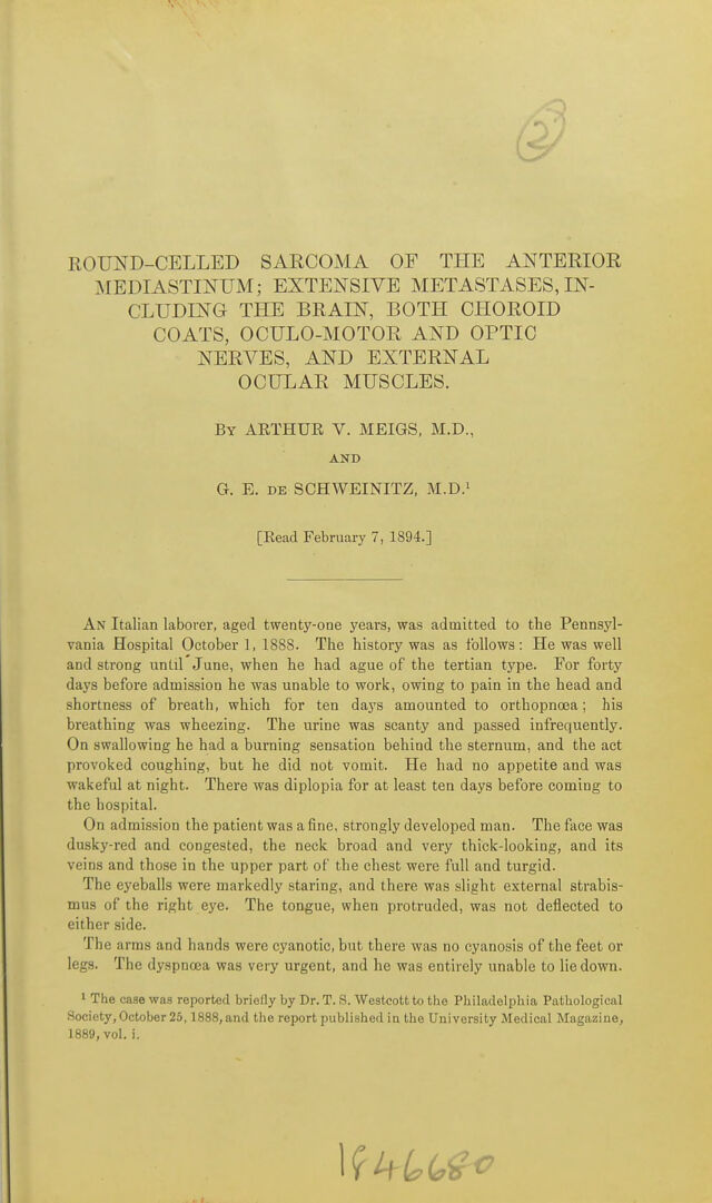
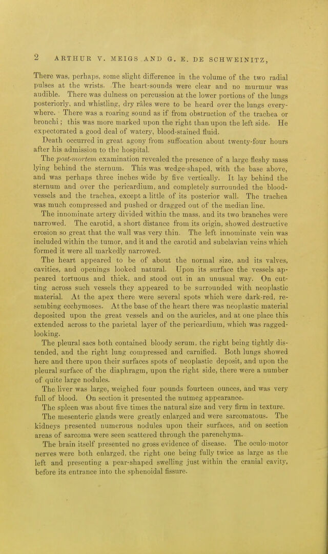
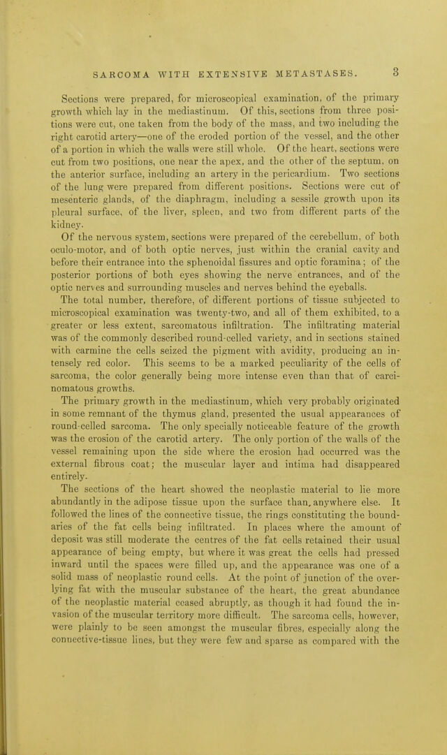
- Arthur V. Meigs
- Date:
- [1894]
Licence: Public Domain Mark
Credit: Round celled sarcoma of the anterior mediastinum : extensive metastases, including the brain, both choroid coats, oculo-motor and optic nerves, and external ocular muscles / by Arthur V. Meigs and G. E. de Schweinitz. Source: Wellcome Collection.
Provider: This material has been provided by UCL Library Services. The original may be consulted at UCL (University College London)
7/16 page 7
![The sclera is free from abnormal appearances, bub in the branches of the ciliary nerves passing through it there are small round cells suggestive of those which have infiltrated the oculo-motor to such a great extent. Posterior half of the left eyeball and 8 mm. of the optic nerve in longi- tudinal section. So far as the optic nerve, optic-nerve entrance, retina, one- half (left) of choroid and sclera are concerned, the description just given applies so nearly that it need not be repeated. The right half of the choroid remains in sitn for 3 mm. from the optic-nerve entrance. Then it becomes detached from the sclera and is greatly thickened. The branching, pig- mented cells are massed together on the outer part, and also surround the well-filled vessels of the stroma, between which are gathered areas of round sarcoma ceils, interspersed with pigment granules and dark, spindle-shaped cells. The chorio-capillaris can be traced unbroken throughout the section, but is indistinct directly over the thickest part of the affected choroidal tissue. The sarcoma cells are dispersed through the layer of choroidal stroma containing large bloodvessels, but are especially massed in several localized areas. Some of the larger veins contain darkly stained cells analogous in their appearance to those which lie outside of the vessel walls. (Fig. 2.) This area of infiltration begins 3 mm. from the nerve entrance, and is ] mm. in diameter at its thickest portion. Transverse section of the contents of the left orbit about 14 mm. anterior to the foramen opttcum.^ The relation of the parts (muscles, vessels, nerves, etc.) one to the other is undisturbed. Muscles. The external, internal, superior rectus, and levator palpebrse superioris are normal in appearance ; the inferior oblique is not included in the section. The inferior lectus contains a patch, constituting about one- third of the section, situated in the upper part, which presents an exquisite picture of infiltration with round sarcoma cells. The connective-tissue in- vestments of the individual fibres and of the fasciculi are literally replaced with the cells of the new growth ; while the sheath of areolar tissue cover- ing the entire muscle in the affected area is crowded with the elements of the neoplasm, which extend into the surrounding fibro-fatty tissue of the orbit. For the most part the diseased area is sharply separated from the portions of the muscle which remain unaffected, although in places small trains of cells proceed a short distance into neighboring areas along the in- ternal perimysium. The section of the superior oblique is more freely supplied with darkly stained nuclei and corpuscles than those of the other muscles, and in one or two spots, in the neighborhood of small bloodvessels, are collections of larger lymphoid cells, probably representing small foci of sarcomatous infil- tration. (Figs. 3 and 4.) ' This section corresponds almost exactly with Table VI. of Lange's Topographische Anatomie des menschlichen Orbital-Inhalts, and has been studied with the aid of this diagram.](https://iiif.wellcomecollection.org/image/b21646375_0009.jp2/full/800%2C/0/default.jpg)


 No text description is available for this image
 No text description is available for this image
 No text description is available for this image