The anatomic histological processes of Bright's disease and their relation to the functional changes : lectures delivered in the Russell Sage Institute of Pathology, City Hospital, New York, during the winter of 1909 / by Horst Oertel.
- Horst Oertel
- Date:
- 1910
Licence: Public Domain Mark
Credit: The anatomic histological processes of Bright's disease and their relation to the functional changes : lectures delivered in the Russell Sage Institute of Pathology, City Hospital, New York, during the winter of 1909 / by Horst Oertel. Source: Wellcome Collection.
Provider: This material has been provided by the Augustus C. Long Health Sciences Library at Columbia University and Columbia University Libraries/Information Services, through the Medical Heritage Library. The original may be consulted at the the Augustus C. Long Health Sciences Library at Columbia University and Columbia University.
118/264 page 94
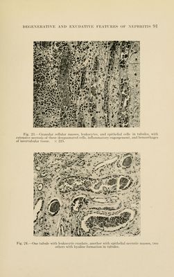
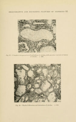
![fuse, the protoplasm disintegrates entirely, and fatty substances tend to appear in the form of fine droplets; vacuoles appear; the lariie granules of the cell, l^y confluence, form necrotic masses, and after ])reaking of the cell membrane, the detritus is dis- charged into the lumen of the tubules (Figs. 21 to 24). In severe cases the lining membrane may be completely desqua- mated (Fig. 28). It is interesting to note, and was first pointed out by Ribbert, that the appearance of fatty substances commences early and primarily in the cells of the loops of Henle, and if we look at such a specimen, one cannot help being astonished at this apparent selective location. Later, however, all the cells under- go the same fate. Changes in the nuclei appear later, l^ut are here of equal severity, leading to their entire loss. They are destroyed either by rapid chromatolysis, with some persistence of the achromatic substance, or the chromatin becomes clumped, solid, and homogeneous, so that the original structure of the nucleus is entirely lost. This condition is known as pyknosis, and eventually results in breaking up into smaller chromatic masses and eventual loss. Finally, nuclei may disappear by a primar}^ peripheral displacement of the normal chromatin masses in the nucleus, leaving its body pale. These chromatin masses are later discharged into the protoplasm of the cell, where they gradually disappear. Proliferation of the epithelium is here a characteristic and important phenomenon. It is excessive, and goes hand in hand with the necrosis of the epithelium and the accumulation of in- flammatory detritus. For this reason it appears usually more prominent in the lower portion of the tubules, where much of the debris, on its removal, stagnates; less so in the upper parts, but may assume there as great dimensions, if the necrosis of the cells and the accumulation of inflammatory detritus assume any proportion](https://iiif.wellcomecollection.org/image/b21211656_0118.jp2/full/800%2C/0/default.jpg)
 No text description is available for this image
 No text description is available for this image
 No text description is available for this image