Half-hours with the microscope : being a popular guide to the use of the microscope as a means of amusement and instruction / illustrated from nature by Tuffen West.
- Tuffen West
- Date:
- [1875?]
Licence: Public Domain Mark
Credit: Half-hours with the microscope : being a popular guide to the use of the microscope as a means of amusement and instruction / illustrated from nature by Tuffen West. Source: Wellcome Collection.
47/124
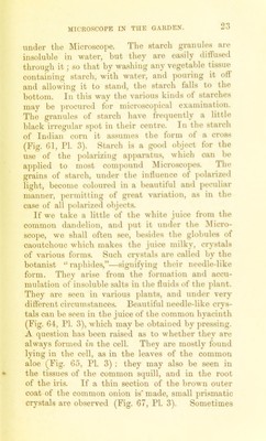
![under the Microscope. The starch granules are insoluble in water, but they are easily diflused through it; so that by washing any vegetable tissue containing starch, with water, and ]>ouring it oti and allowing it to stand, the starch falls to the bottom. In this way the various kinds of starches may be procured for microscopical examination. The granules of stai'ch have frequently a little black irregular spot in their centre. In the starch of Indian corn it assumes the form of a cross (Fig. Gl, ri. 3). Starch is a good object for the u.se of the j)olariziug apparatus, which can be applied to most compound Microscopes. The grains of starch, under the intluence ot jiolarized light, become coloured in a beautiful and peculiar manner, permitting of great variation, as in the case of all polarized objects. If we take a little of the white juice from the common daiulelion, and put it under the Micro- scope, we shall often see, be.sides the globules of caoutchouc which makes the juice milky, crystals of various forms. Such crystals are called by the botanist “ raj)hides,”—signifying their needle-like form. They arise from the formation and accu- mulation of insoluble salts in the fluids of the ])lant. They are seen in various plants, and under very dirterent circumstances. Beautiful needle-like crys- tals can be seen in the juice of the common hyacinth (Fig. 64, PI. 3), which may be obtained by pressing. A question has been raised as to whether they are always formed in the cell. They are mostly found lying in the cell, as in the leaves of the common aloe (Fig. 65, PI. 3) : they may also be seen in the tissues of the common squill, and in the root of the iris. If a thin section of the brown outer coat of the common onion is made, small prismatic crystals are observed (Fig. 67, PI. 3). Sometimes](https://iiif.wellcomecollection.org/image/b28099436_0047.jp2/full/800%2C/0/default.jpg)