Philips' anatomical model : a pictorial representation of the human frame and its organs, with descriptive text / by Dr. Schmidt ; English edition by William S. Furneaux.
- Eduard Oscar Schmidt
- Date:
- [1893?]
Licence: Public Domain Mark
Credit: Philips' anatomical model : a pictorial representation of the human frame and its organs, with descriptive text / by Dr. Schmidt ; English edition by William S. Furneaux. Source: Wellcome Collection.
8/22
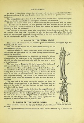
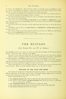
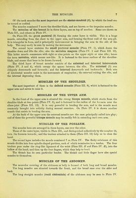
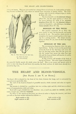
![of which is the coccyx (C). These all form together a basin-like cavity, the pelvis, in which the organs of the abdomen are supported. There is a socket on the lower outside portion of each hip-bone, in which the upper rounded extremity of the thigh-bone moves. The thigh-bone (/), which is very strong, has the upper part bent at an angle to the lower; and on this the rounded head, forming the joint, is situated. The lower part terminates in two knobs (hi, Fig. 1), partially covered with gristle, with which the bones of the calf are articulated. These bones are called the tibia or shin-bone {t) and the fibula or splint-bone (/), and form, with the thigh-bone, the knee-joint, which is protected in front by the little round knee- cap or patella (pa). The lower ends of the shin-bone and the splint-bone form the joint to which the foot is attached. The foot is composed of seven tarsal bones, five metatarsal or instep bones (m/), and the bones of the toes. The largest of the tarsal bones projects backward and forms the heel (ca), and the top one (to) forms the ankle-joint with the two calf-bones. The big toe, like the thumb, has two bones; while the other’s have three each. THE MUSCLES. [See Plates III. and IY. of Model.] The muscles are the active organs, by means of which the movements of the human body are effected. They form what is known as the flesh. By means of the nerves, by which they are controlled, they may be contracted or relaxed. Muscles consist of bundles of fibres, covered with a whitish skin or membrane. The ends of a muscle terminate in tendons, by means of which they are fastened to the bones. As the muscles have a great deal of work to perform, they rapidly waste away; therefore they are pai’ticulaily well provided with blood-vessels, by means of which the used-up particles are removed, and fresh nourishment supplied to make good the loss. IMost of the muscles are in the shape of spindles ; but some, such as those on the breast, are broad and flat. They are mostly placed in layers, one on the top of another. MUSCLES OF THE FACE AND HEAD. The muscles of the face have two important functions to perform : viz., the formation of the different expressions of the face, such as those of laughter, crying, anger, joy, etc.; and the control of the movements of the mouth, eye-lids, nose, etc. Those of special interest are:— 1. The temporal muscle (Plate III. 1), which lifts the lower jaw in the act of biting. 2. The muscle of the forehead (occipito-frontalis, Plate III. 2), by means of which the brow is contracted or wrinkled. 3. The orbicularis muscle (3), by which the eye-lids are closed. 4. The muscles which raise the upper lip and control the corners of the mouth (4)](https://iiif.wellcomecollection.org/image/b28128679_0008.jp2/full/800%2C/0/default.jpg)