On the difference in the mode of ossification of the first and other metacarpal and metatarsal bones / by Allen Thomson.
- Allen Thomson
- Date:
- [1868?]
Licence: Public Domain Mark
Credit: On the difference in the mode of ossification of the first and other metacarpal and metatarsal bones / by Allen Thomson. Source: Wellcome Collection.
Provider: This material has been provided by The University of Glasgow Library. The original may be consulted at The University of Glasgow Library.
17/20
![at the proximal, or carpal, ends of the metatarsal hones of the pollex and hallux. The cartilaginous lines separating the distal epiphyses from the shafts are cpiite visible; though they have been to some extent traversed by ossification near the centre. There is also an epiphysis at the proximal end of the second, or index, metacarpal, though the cartilaginous line separating it from the shaft is not quite so clear, especially towards the centre of the bone, as it is in the distal ends of the metacarpals of the pollex and hallux. There are no visible traces of epiphysial lines in the proximal end.s of the other metacarpals or in the .5th metatarsal. The three middle metatarsals have not l>een cut; ami the presence or al)s**nce ot epiphysial lines cannot therefore be determined in them. G. iL Humphry.] DESCRIFFION OF THE PLATE. In the several figures the numlx^rs ami letters indicat*? details as follows, viz. I, the first iuctacaii»al or meUitarsal bone™ II, the second ditto. 1, 2, 3, tl>e proximal, middh* and terminal phalanges, in all ex*?epting Fig. 6, in which there are seven phalanges marked in the middle digit. p, the more common or usual pro.ximal epiphyses; p\ the less common and sometimes im|)erfect proximal epiphyses. d, the m*>ro comuion or usual distal ejiiphyses; d\ the leas common and sometimes imperfect distid epiphyses. In a numl)er of the figures a slightly curved line marke<l f, repre- sents tlie direction of a bristle intrmluce*! into the csuml of the Ixmes througli the so-callwl nutritious foramen; that dii-ection being in most instances towards the extremity which is soonest ossified, or in which an epiphysis is wanting. Fig. 1. Dorsal view- of the dried metacarpal and j)lmlangeal lones of the first and second fingers (thumb and forefinger) from the left hand of a girl of eight years of age : d\ the fissure separating a *listal ejiiphysis on the first mefiicarpal bone; p\ a small but ileep fissure which indicates a partml proximal epiphysis on the second inetacuqml bone. Fig. 2. Antero posterior longitudinal section of the first and second metacar|)al bones and digital phalanges of the thumb and forefinger from the left hand of a child of seven years of age in the VOL III. 10](https://iiif.wellcomecollection.org/image/b24931445_0017.jp2/full/800%2C/0/default.jpg)


 No text description is available for this image
 No text description is available for this image
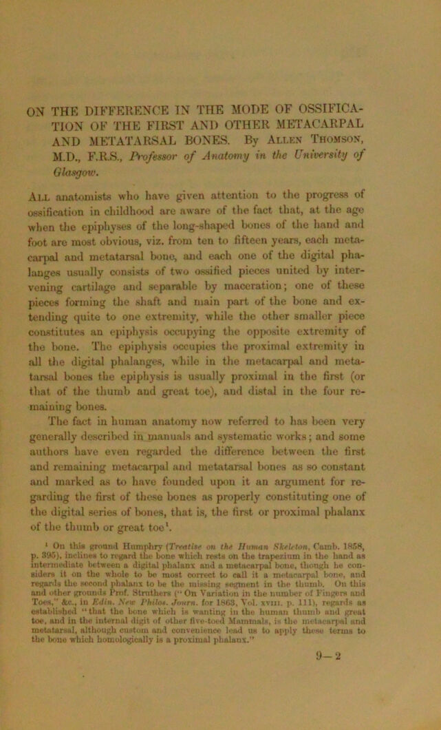 No text description is available for this image