Anatomico-chirurgical observations on dislocations of the astragalus / by Thomas Turner.
- Turner, Thomas, (1793-1873)
- Date:
- 1843
Licence: Public Domain Mark
Credit: Anatomico-chirurgical observations on dislocations of the astragalus / by Thomas Turner. Source: Wellcome Collection.
Provider: This material has been provided by The Royal College of Surgeons of England. The original may be consulted at The Royal College of Surgeons of England.
135/148 (page 129)
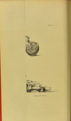
![< former bones should be drawn towards the latter, as seen in plate 1; but, secondly, if the astragalus be partially displaced outwards and forwards, n and fractured, and one or more of the fragments ] of the bone should remain interposed between i the cup of the os naviculare and the anterior i inferior edge of the tibia, that this bone would be still drawn downwards, or the foot upwards, j but its articular surface would occupy the posterior ■ astragalar facet of the os calcis, instead of the ii anterior; thus, the posterior edge of the tibia t! recedes, or is situated nearer the tuberosity of the heel than when the astragalus is wholly luxated or excised ; and the heel in this case will be shortened i instead of lengthened, as it appears to be when the ; hollow of the os calcis has been vacated by the Is astragalus, and gives occupancy to the tibia, which y descends upon it. If plate 3 be compared with plate 1, it will be noticed, that in the former instance, as in the latter, the malleolar process of the tibia is closely applied 5 to the os calcis, but in the first case the bone is t anchylosed with it. The apposition of the tibia to 1 the os calcis is complete at the malleolus interims ; uj but in the front view of the preparation, from fj which the engraving is taken, the position of the line of the under articulating surface is oblique | from within outwards, the obliquity inclining out- wards and upwards. This is caused by a portion of the fractured astragalus being interposed, and an- chylosed to the tibia, fibula, and calcaneum. The fibula and outer edge of the under articulating sur- face of the tibia being thus mechanically prevented](https://iiif.wellcomecollection.org/image/b22348281_0137.jp2/full/800%2C/0/default.jpg)