Volume 1
A system of surgery / by Charles B. Ball [and others] ; edited by Frederick Treves.
- Date:
- 1896-1898
Licence: Public Domain Mark
Credit: A system of surgery / by Charles B. Ball [and others] ; edited by Frederick Treves. Source: Wellcome Collection.
Provider: This material has been provided by King’s College London. The original may be consulted at King’s College London.
1151/1206 (page 1109)
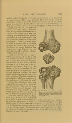
![position for several hours, also to check oozing. The result is almost invariably primary union, without a trace of suppuration. The stitches are taken out on tlie ninth or tenth day, and all is sound. Of course a careful watch is kept upon the temperature all this time, and if this rises and remains elevated and the patient complains of pain and throbbing in the joint with malaise and anorexia, the dressings must be removed and the joint inspected. If it be found distended with either blood or pus, a tube must be inserted and thorough drainage provided for. In dissecting away the synovial membrane I always commence methodically at the tip of the oval flap when it is turned up, and work upwards, peeling it from the posterior surface of the quadriceps, then peel it off from the point of the femur and at the sides, then from about the crucial ligaments, and, finally, off the head and borders of the tibia. In many cases it is possible in this way to remove the whole synovial membrane almost in a continuous sheet. If the patellar ligament has been divided I always stitch it together again with silk, which remains buried. It is well in these operations not to include the ca])sule of the joint in the stitches which unite the skin wound. If the capsule and ex])ansion of the quadriceps be included, the skin is apt to be dragged in and the edges displaced, which may interfere with primary union. Indeed, it is probably better, for sevei’al reasons, not to unite the edges of the capsule and the fibrous expansion of the muscle at all. One advantage of not doing so is that, if there be any effusion into the joint cavity after the operation, the fluid is not pent up, but escapes into the areolar planes around the knee, and is more rapidly absorbed than if it remained in the joint. And should the deeper parts unhappily suppurate from any mischance, the pus would at once appear under the skin, and be easily evacuated, which would not be the case were the divided capsule closely sutured. When the wound is perfectly healed, which, as a rule, is the case at the end of ten days, I find it best to put up the whole limb, from the gluteal fold to the toes, in a firm plaster-of-Paris case^ applied closely over a thin layer of cotton wadding. This splint can be removed at the end of six weeks, and renewed if the joint is not yet firm enough to walk upon. As a rule, the limb may be used in the eighth week, but not without the support of the sjjlint. In some cases, however, where only a very limited removal of synovial mem- brane has been undertaken, and where all the more important ligaments have been spared, a movable joint may be aimed at, and here it will be necessary to leave off the splints during the day,' and to encourage passive and active movement. Massage, too, should be employed to get rid of the stiffness which remains. But in these cases it will be very desirable to keep the limb on a straight splint at night, lest angular displacement take place, which would be difficult to correct during the day. But, hitherto, the author has only operated, a.s a rule, upon such cases as were affected with too much disease to justify the hope that after operation the movements](https://iiif.wellcomecollection.org/image/b21303691_0001_1151.jp2/full/800%2C/0/default.jpg)