Repeated paroxysmal failure of sight in connection with heart disease / by Edward Nettleship.
- Edward Nettleship
- Date:
- [1879]
Licence: Public Domain Mark
Credit: Repeated paroxysmal failure of sight in connection with heart disease / by Edward Nettleship. Source: Wellcome Collection.
Provider: This material has been provided by UCL Library Services. The original may be consulted at UCL (University College London)
2/8 page 2
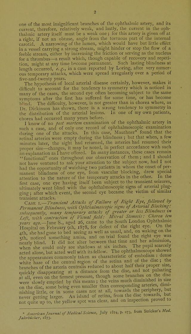
![one of the most insignificant branches of the ophthalmic arter)', and its current, therefore, relatively weak, and lastly, the current in the oph- thalmic artery itself must be a weak one ; for this artery is given otTat a right, if not an obtuse, angle from the tenuous part of the internal carotid. A narrowing of the lumen, which would have but httle effect in a vessel carrying a strong stream, might hinder or stop the flow of a feeble stream, either by increasing the friction or serving as the nucleus for a thrombus—a result which, though capable of recovery and repeti- tion, mit'ht at any time become permanent. Such lasting blindness at leng'th occurred, e.g., in a case reported by Loring, after very numer- ous'^temporary attacks, which were spread irregularly over a period of five-and-twenty years. The hypothesis of local arterial disease certainly, however, makes it diflicult to account for the tendency to symmetry which is noticed in many of the cases, the second eye often becoming subject to the same symptoms after the first has suffered for some time, or has become blind. The difficulty, however, is not greater than in chorea where, as ]Jr. Dickinson has shown, there is a strong tendency to symmetry in the distribution of the arterial lesions. In one of my own patients, chorea had occurred many years before. I know of no post mortem examination of the ophthalmic artery_ m such a case, and of only one record of ophthalmoscopic examination during one of the attacks. In this case, Mauthner* found that the retinal arteries were empty during the blindness ; but that when, a few minutes later, the sight had returned, the arteries had resumed their |)roper size changes, it may be noted, in perfect accordance with such an explanation as I have offered. In many instances, these cases remain functional ones throughout our observation of them; and I should not have ventured to ask your attention to the subject now, had I not had the opportunity of watching two patients in whom the onset of per- manent blindness of one eye, from vascular blocking, drew special attention to the nature of the temporary attacks in the other. In the first case, one eye having itself been subject to the transient seizures, ultimately went blind with the ophthalmoscopic signs of arterial plug- ging ; after which event, the second eye became the victim of similar transient attacks. , „. , ^ ' j-„ j , Case \.—Transient Attacks of Failure of Right Eye, followed by Permanent Blindness, with Ophthalmoscopic signs of Arterial Blocking: subsequently, many temporary attacks of greater or less Blutdness in Left, with contraction of Visual field: Mitral Stenosis: Chorea tai years Jane S., aged 26, came to the South London Ophthalmic Hospital on February 9th, 1878, for defect of the right eye. On the 4th she had gone to bed seeing as well as usual, and, on \yaking on the Sth, noticed something amiss, and on trial found the right eye was nearly blind. It did not alter between that time and her admission, when she could only see shadows at six inches. The pupil scarcely acted alone, but acted well with its fellow. The ophthalmoscope showed the appearances commonly taken as characteristic of embolism : dense white haze of the central region of the retina and of the disc; the branches of the arteria centralis reduced to about half their proper size, quickly disappearing at a distance from the disc, and not pulsating at all, even on the firmest pressure, though some branches on the disc were slowly emptied by this means ; the veins much diminished m size on the disc, some being even smaller than corresponding arteries, dimi- nishing little, or in some cases not at all, towards the periphery, but never getting larger. An island of retina, from the disc towards, but not quite up to, the yellow spot was clear, and on inspection proved to • American Journal of Medical Science, July .8,4, p. 273, from Su-icker's Mrrf. JakrlrSclier, 1873.](https://iiif.wellcomecollection.org/image/b21645218_0004.jp2/full/800%2C/0/default.jpg)