A text-book of the diseases of the ear and adjacent organs / by Dr. Adam Politzer ; translated from the third German edition by Oscar Dodd ; edited by Sir William Dalby.
- Ádám Politzer
- Date:
- 1894
Licence: Public Domain Mark
Credit: A text-book of the diseases of the ear and adjacent organs / by Dr. Adam Politzer ; translated from the third German edition by Oscar Dodd ; edited by Sir William Dalby. Source: Wellcome Collection.
Provider: This material has been provided by the Harvey Cushing/John Hay Whitney Medical Library at Yale University, through the Medical Heritage Library. The original may be consulted at the Harvey Cushing/John Hay Whitney Medical Library at Yale University.
24/764 page 6
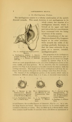
![With the progressing growth of the cranial bones in the early years of life, the following alterations take place in the squamous and tympanic portion-. While (Fig. 7) the superior part of the squamous portion is placed on the Mr Fig. Fig /' g 7.—Temporal Bone in the New-born Infant. Superior part of the squamous bone ; b, Its inferior part below the line of the zygomatic process ; c, Annulus tympanicus ; d, d, The fissure between squamous portion and mastoid process, reaching to the foramen stylo-mastoi- deum : e, Foramen stylo-mastoideum ;J] Fora- men ovale : gt Foramen rotundum (left ear). Literal part of the cranium, its lower portion (6). which lies beneath the line of the zygomatic process, takes a more horizontal position, in such a way that in the completely developed temporal bone the superior part of the squamous portion (Fig. 9. a) is bent almost at a right angle to its inferior horizontal portion (b). This horizontal portion forms the superior wall of the osseous meatus, and in con- junction with the mastoid process it also forms a part of the posterior wall. As mentioned above, an essential part in this forma- tion of the osseous meatus is taken by the tympanic por- tion of the temporal bone. With its growth, through deposit of osseous substance on its exterior (Zuckerkandl), there arises an osseous groove (Fig. 8. 6), the lateral walls of which reach so far up in a median direction near to the tympanic bone, that they also take part to a varying extent in the formation of the superior wall of the meatus. c b Fig. 8.—Osseous Meatus in the Adult. a, Horizontal part of the squamous bone (superior part of the meatus) ; b, Tympanic portion ; c, Lumen of the meatus ; d, Mastoid process (left ear).](https://iiif.wellcomecollection.org/image/b21007883_0024.jp2/full/800%2C/0/default.jpg)