Licence: Public Domain Mark
Credit: Practical anatomy. Source: Wellcome Collection.
Provider: This material has been provided by the Harvey Cushing/John Hay Whitney Medical Library at Yale University, through the Medical Heritage Library. The original may be consulted at the Harvey Cushing/John Hay Whitney Medical Library at Yale University.
61/600
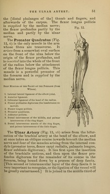
![Fig. 12. the [distal phalanges of the] thumb and fingers, and afterwards of the carpus. The flexor longus pollicis is supplied by the median nerve; the flexor profundus partly by the median and partly by the ulnar nerve. The Pronator Quadratus (Fig. 12, 6) is the only muscle in the arm whose fibres are transverse. It arises from a somewhat oval surface on the front of the ulna below the origin of the flexor profundus, and is inserted into the wThole of the front of the radius below the attachment of the flexor longus pollicis. This muscle is a powerful pronator of the forearm and is supplied by the median nerve. Deep Muscles of the Front of the Forearm (from Wilson). 1. Internal lateral ligament of the elbovr-j oint. 2. Anterior ligament. 3. Orbicular ligament of the head of the radius. 4. Flexor profundus digitorum (the lumbricales re- moved). 5. Flexor longus pollicis. 6. Pronator quadratus. 7. Adductor pollicis. 8. Dorsal interosseous of the middle, and palmar interosseous of the ring fiuger. 9. Dorsal interosseous muscle of the ring finger, and palmar interosseous of the little finger. The Ulnar Artery (Fig. 13, 16) arises from the bifur- cation of the brachial artery at the bend of the elbow, and at once takes an oblique course inwards beneath the median nerve and four of the muscles arising from the internal con- dyle (pronator teres, flexor carpi radialis, palmaris longus, flexor sublimis digitorum). It lies first upon the insertion of the brachialis anticus, and then upon the flexor pro- fundus digitorum for the remainder of its course in the forearm, being bound down by a process of deep fascia. [This second or intermuscular layer of the deep fascia it is veiy important to recognize, or the ligation of the vessel will be greatly embarrassed.] It is j oined in the middle third of](https://iiif.wellcomecollection.org/image/b21020735_0061.jp2/full/800%2C/0/default.jpg)