Licence: Public Domain Mark
Credit: Practical anatomy. Source: Wellcome Collection.
Provider: This material has been provided by the Harvey Cushing/John Hay Whitney Medical Library at Yale University, through the Medical Heritage Library. The original may be consulted at the Harvey Cushing/John Hay Whitney Medical Library at Yale University.
64/600
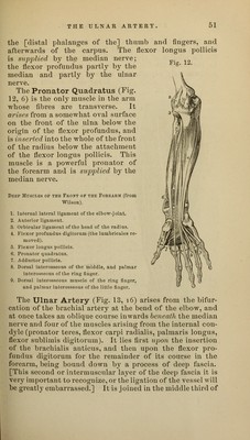
![the front and back of the carpus, and anastomose with corresponding branches from the radial.1 The Median Nerve (Figs. 11 and 13, pp. 4? and 52) enters the forearm between the heads of the pronator teres, and then passing between the origins of the flexor sublimis digitorum, crosses the ulnar artery to lie between the flexor sublimis and flexor profundus muscles. It is placed superficially between the flexor tendons near the wrist, and passes with them beneath the annular ligament into the hand. Branches.—The median nerve supplies all the muscles of the front of the forearm except the flexor carpi ulnaris and half the flexor profundus, in the following manner: As soon as it enters the forearm it gives branches to the pronator teres, flexor carpi radialis and flexor sublimis digitorum, and, after crossing the ulnar artery, gives off the anterior interosseous nerve (Fig. 13, 25). This passes down the front of the interosseous membrane, giving branches to the flexor loligus pollicis and the outer half of the profundus digitorum, and then beneath the pronator quadratus (which it supplies) to the front of the wrist- joint, where it gives a branch to the articulation. A cutaneous palmar branch (Fig. 11, 30) arises a short distance above the annular ligament, over which it passes to be distributed to the skin of the palm. The Ulnar Nerve (Fig. 13, 2, 2) enters the forearm behind the internal condyle, by passing between the heads of the flexor carpi ulnaris. It lies under cover of that muscle and upon the flexor profundus digitorum for the whole of its course in the forearm ; and about the middle [third] of the [fore]arm comes into close relation with the ulnar artery, and, keeping to the ulnar side [see rule, p. 48], accompanies it over the annular ligament into the palm. Branches (Fig. 13).—The ulnar nerve gives small articular branches to the back of the elbow, and supplies one and a half of the muscles of the forearm, viz., the flexor carpi ulnaris and the inner half of the flexor pro- fundus digitorum. In the lower third of the forearm the nerves give a dor- 1 Professor Ellis enumerates a metacarpal branch which is usually the continuation of the posterior carpal artery to the back of the 5th metacarpal bone, as will be seen in the dissection of the back of the hand.](https://iiif.wellcomecollection.org/image/b21020735_0064.jp2/full/800%2C/0/default.jpg)