Licence: Public Domain Mark
Credit: Practical anatomy. Source: Wellcome Collection.
Provider: This material has been provided by the Harvey Cushing/John Hay Whitney Medical Library at Yale University, through the Medical Heritage Library. The original may be consulted at the Harvey Cushing/John Hay Whitney Medical Library at Yale University.
69/600
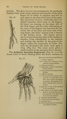
![the wrist, and is only a thickened portion of the common fascia of the limb. It is attached to the scaphoid bone and to the ridge of the trapezium on the outer side, and to the unciform process of the unciform bone and slightly to the pisiform bone on the inner side [that is, to the first and last bones of each row.] The ulnar artery and nerve, and the cutaneous palmar nerves cross it, and also the tendon of the palmaris longus in part. (The tendon of the flexor carpi ulnaris sends an expansion over the ulnar artery and nerve, which must not be mistaken for the annular ligament itself.) When divided in the middle, it will be seen to be perforated by the tendon of the flexor carpi radialis, and to have beneath it the median nerve and the tendons of the flexors of the thumb and fingers. A quantity of loose bursal tissue will be found beneath the annular ligament and closely connected with the tendons and median nerve ; its use is to facilitate the movements of the tendons, and it occasionally becomes diseased, when fluid is developed in it in considerable quantity, and forms a fluctuating tumor above and below the annular ligament, often con- taining numerous rice-like bodies. This tissue must be carefully dissected away. [Like the sheaths of the flexor tendons and the palmar fascia, this also acts as a pulley.] The Median Nerve (Fig. 13, 3.3, p. 52) passes beneath the annular ligament superficially to the tendons, and di- vides into two trunks which subdivide into four digital nerves. The first or outermost, after giving a small branch to supply some of the short muscles of the thumb, bifur- cates into branches to supply the two sides of the palmar aspect of the thumb; the second digital nerve supplies the radial side of the index finger after giving a small twig to the first lumbricalis muscle ; the third, after supplying the second lumbricalis, bifurcates near the root of the finger to supply the ulnar side of the index and the radial side of the middle fingers; the fourth bifurcates to supply the ulnar side of the middle finger and the radial side of the ring finger, this last branch joining the branch from the ulnar nerve previously seen. The ulnar artery is to be divided beyond the origin of the profunda and the arch turned down as far as possible, but the nerve may be left uncut. The median nerve is to be divided at the wrist and turned down, and the flexor tendons with the lumbricales muscles](https://iiif.wellcomecollection.org/image/b21020735_0069.jp2/full/800%2C/0/default.jpg)