Licence: Public Domain Mark
Credit: Human embryology / by Charles Sedgwick Minot. Source: Wellcome Collection.
47/854 page 15
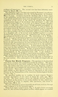
![enlarged with eertain accompanying modifications to obtain the tissue figured in Fig. 7. There is no special formation of cells around the blood-vessels, where, according to Ercolani, the decidual tissue arises by new formation. In Turner's specimens the upper part of the compact layer was imperfectly preserved, but according to his description there appears to have been a coagulum similar to that which I have found, but thicker. In the deep part of the layer the cells are less enlarged, and when the cavernous layer is reached, there occurs a rapid transition in the character of the cells, which become smaller and more fusiform, and their nuclei more elongate, smaller, and deeper stained by alum-cochineal. The gland openings upon the surface of the uterus lead into tubes. Fig. 6, gZ', which run slightly obliquely through the compact layer, taking a more or less nearly straight course and joining the contorted gland tubes. Fig. 6, gl\ of the cavernous layer. The gland ducts are completely devoid of lining epithe- lium, which has cnag]. '^^^^^.f '^T-^^'f-^^^^'^y^A'^ . disappeared ex- cept for a very loose cell, occa- sionally found lying free in the ducts; the cells have not fallen out from the sec- tions, but were lost before the tissue was im- bedded.* The ducts then are wide tubes run- ning nearly straight through the upper part of the decidua and bounded direct- ly by the decid- ual tissue; they c o m m u n icate below with the contorted cavi- ties. _ . .__J The cavern- Fig. r.—uterus cue mouth iji-eguaut; pui-tiou or the coiiipcict layer rkn« la^m-pd onn of the decidua seen in vertical section; coagl, coagulum upon the sur- uute la^eib cun- face; d, d', decidual cells, x 445 diams. tain numerous spaces, the areolge of Turner, 79.1, 547, who was uncertain as to their character, though he ascertained that many of them belong to the glandular system. In my specimen it is perfectly clear that all the larger areolae belong to the glands, which must be extremely dis- torted and distended to give the shapes shown in Fig. 6. The thin * The blocks to be cut were stained in toto with alum-cochineal and eosin, imbedded in paraf- fin, etc. The sections were fastened on the side with celloidin, to keep the parts m place.](https://iiif.wellcomecollection.org/image/b20412162_0047.jp2/full/800%2C/0/default.jpg)


 No text description is available for this image
 No text description is available for this image
 No text description is available for this image