Licence: Public Domain Mark
Credit: Practical manual of obstetrics. Source: Wellcome Collection.
Provider: This material has been provided by the Harvey Cushing/John Hay Whitney Medical Library at Yale University, through the Medical Heritage Library. The original may be consulted at the Harvey Cushing/John Hay Whitney Medical Library at Yale University.
45/430 page 15
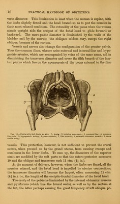
![At about weaning time (la deuxitme enfance) the characteristics just mentioned disappear, and in the third year of life the differences in the pelvis of the two sexes are quite marked ; but it is not until the seventeenth year that the pelvis attains its full size, and the three bones of the os in- nominatum become united. [Child-bearing in women under twenty years of age quite commonly leads to subsequent pelvic breadth, producing a marked change in the female figure. This can be accounted for upon the supposition that complete ossification and full pelvic size is not attained until about that year.—Ed.] The pelvis measurements viewed generally are smaller in the human species than in other animals, and the pelvis of the human male is, gener- ally, smaller than that of the human female. Studying the lower species (mammalians) the sacro-vertebral angle no longer exists, save in the monkey, which animal has much in common with man ; the sacrum is almost on a line with the vertebral column. The two innominate bones are very close together, and though long drawn out they only form a kind of non-excavated ring ; the two straits have one and the same axis, forming, with the axis of the body, almost a straight line ; and it is on account of this and the more or less elongated head of the foetus in mammalians that delivery is so much easier in other vertebrate animals than in the human species. I cannot close this study of the differences of the pelvis without men- tioning—were it solely for its originality—a classification of mammalians according to their mode of delivery proposed by Joulin. It might be called the obstetrician's classification. 1. Pre-ischiatic deliveries. The foetus takes its exit in front of the is- chial tubers. The human female is the one and only example. 2. Inter-ischiatic deliveries. The foetus comes out between the tuber- osities of the ischia. The large guenons (pouched monkeys), opossums, and cabiai. 3. Post-ischiatic deliveries. This class is the most numerous, embrac- ing animals baring a short sacrum : domestic animals, mares, cows, and deer. Art. IV.—Difference Due to the Presence of the Soft Pai: The soft parts lining the pelvic cavity modify to a greater or Leas extent its form and dimensions. In the greater pelvis the anterior wall—wanting in the skeleton—is made up of the abdominal muscles. The internal iliac fossa is tilled up by the iliacus and psoas, whose presence slightly niters the form of the superior strait, which, still resembling a triangle with rounded corners, has now its base in front and apex behind. The psoas muscles which run aa from the spine to Poupart's ligament diminish, by 2 ctm. (] in. |, the tran>-](https://iiif.wellcomecollection.org/image/b2100013x_0045.jp2/full/800%2C/0/default.jpg)