Licence: Public Domain Mark
Credit: Practical manual of obstetrics. Source: Wellcome Collection.
Provider: This material has been provided by the Harvey Cushing/John Hay Whitney Medical Library at Yale University, through the Medical Heritage Library. The original may be consulted at the Harvey Cushing/John Hay Whitney Medical Library at Yale University.
69/430 page 39
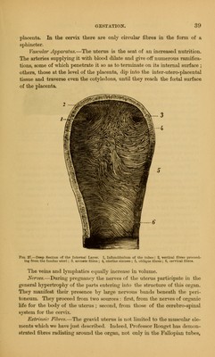
![placenta. In the cervix there are only circular fibres in the form of a sphincter. Vascular Apparatus.—The uterus is the seat of an increased nutrition. The arteries supplying it with blood dilate and give off numerous ramifica- tions, some of which penetrate it so as to terminate on its internal surface ; others, those at the level of the placenta, dip into the inter-utero-placental tissue and traverse even the cotyledons, until they reach the foetal surface of the placenta. Fig. 27.—Deep Section <>f the Internal Lnyor. 1, Infuinlibuhun of the mhos : 2, Yortio:il fibro- ]>n>.- 1- intf from the fundus uteri ; .'S. Annate tiling ; 1, uterine Mini>e~ ; .'.. obllqtM rvu-.il libres. The veins and lymphatics equally increase in volume. Nerves.—During pregnancy the nerves of the uterus participate in the general hypertrophy of the parte entering into the structure of this organ. They manifest their presence by large nervous bands beneath the peri- toneum. They proceed from two sources : first, from the nerves of organic life for (he body of the uterus ; second, from tlm-^c of the cerebrospinal system for the cervix. Extrinsi-- Fibres, The gravid Uterus is not limited to the muscular ele- ments which we have just described. Indeed. Professor EtoUgel has demon- strated fibres radiating around the organ, not only in the Fallopian tubes,](https://iiif.wellcomecollection.org/image/b2100013x_0069.jp2/full/800%2C/0/default.jpg)