The science and art of obstetrics / by Theophilus Parvin.
- Theophilus Parvin
- Date:
- 1895
Licence: Public Domain Mark
Credit: The science and art of obstetrics / by Theophilus Parvin. Source: Wellcome Collection.
83/693 page 82
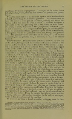
![PHYSIOLOG Y OF PBE( 1 \. i N( ) Fig.47. longitudinal fold of the germinative epithelium which is sunk in the con nective tissue of the external lateral part of the Wolffian body ; this folc is covered over and thus converted into i tube. It remains open externally, anw that portion becomes the pavilion. Tin occurrence of secondary pavilions, to bo referred to in the description of the ovii duct, is readily explained, according frt Duval, by supposing that the canal o Muller is not completely closed in all iti extent at the period of embryonic lifi when the two borders of the gutter, whic:' give origin to it, are turned toward eaci other, in order to transform the guttet into a canal. Miiller's ducts unite in part of their course to form the uterui and the vagina ; the limit to this union i the insertion of the round ligament, whic is the analogue of the gubernaculum testt in the male, and has the same relatioi with the inguinal canal. All of the du< below the round ligament unites with tl corresponding portion of the duct on tl other side, thus making at first^ a doub uterus and vagina ; but absorption of ttl intervening wall occurs, and each orgs; is single. The left duct is usually fu ther in front than the right, and the tv are united in this oblique position; tl presence of the intestine upon the left side is thought to explain tl fact that the left duct is placed further to the front than the ngt The fusion of the two ducts is complete in the embryo of two montl The point at which this union begins is unsettled. Kolliker believ that it is at the middle of Miiller's canals, while others hold that takes place from below upward. All that portion of the duct abo the insertion of the round ligament becomes the oviduct. The pron nence on the internal face of the Wolffian body is composed of a ran of embryonic connective tissue covered by well-developed germ epitb Hum; it is the first rudiment of the genital gland, and is found all in the embryo which is to be a male, as well as in that which is to be female; in the female the ovaries and ovules are derived from the c] thelial covering, while the outgrowth itself is destined to furnish t vascular stroma of the ovary. The next change that is observed is the appearance of cells, whi are round, have a well-developed nucleus and distinct nucleolus; thm are the primordial ovules or primitive ova. At the deep part oi U genital eminence, and in close contact with it, sections show that 11 tubes of the upper portion of the Wolffian body are narrower ami m a clearer epithelium than those of the lower portion; the supenoi known as the sexual or genital portion, the inferior as the urinary. Diagrammatic Outline op the Wolffian Bodies, and their Re- lation to the Ducts of Muller and the Reproductive Glands. (From Allen Thomson.) ot. Seat of origin of ovary or testes. W. Wolffian body. w. Wolffian duet. m. Duct of Muller. gc. Genital cord. ug. Dro-genital sinus, i. Rectum, cl. Cloaca.](https://iiif.wellcomecollection.org/image/b20386394_0084.jp2/full/800%2C/0/default.jpg)



 No text description is available for this image
 No text description is available for this image
 No text description is available for this image