A text-book of human physiology : including histology and microscopical anatomy, with especial reference to the practice of medicine.
- Landois, Leonard
- Date:
- 1904
Licence: In copyright
Credit: A text-book of human physiology : including histology and microscopical anatomy, with especial reference to the practice of medicine. Source: Wellcome Collection.
Provider: This material has been provided by The University of Leeds Library. The original may be consulted at The University of Leeds Library.
66/1086 page 48
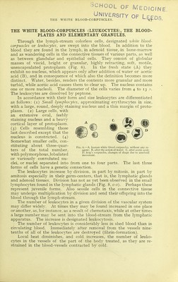
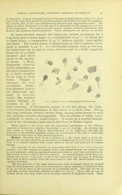
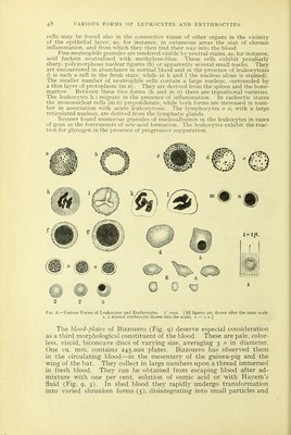
![cells may be fotind also in the connective tissue of other organs in the vicinity of the epithelial layer, as, for instance, in cutaneous areas the seat of chronic inflammation, and from which they then find their way into the blood. Fine neutrophile granules are rendered visible by neutral stains, as, for instance, acid fuchsin neutralized with methylene-blue. These cells exhibit peculiarly sharp, polymorphous nuclear figures (h) or apparently several small nuclei. They are encountered in abundance in normal blood and in the presence of leukocytosis (i is such a cell in the fresh state, while in k and 1 the nucleus alone is stained). The smaller number of neutrophile cells contain a large nucleus, surrounded by a thin layer of protoplasm (m n). They are derived from the spleen and the bone- marrow. Between these two forms (h and m n) there are transitional varieties. The leukocytes h i migrate in the presence of inflammation. In cachectic states the mononuclear cells (m n) preponderate, while both forms are increased in num- ber in association with acute leukocytosis. The lymphocytes o o, with a large reticulated nucleus, are derived from the lymphatic glands. Neusser found numerous granules of nucleoalbumin in the leukocytes in cases of gout as the forerunners of uric-acid formation. The leukocytes exhibit the reac- tion for glycogen in the presence of progressive suppuration. 2 2 S Fig. 8.—Various Forms of Leukocytes and Erythrocytes. X looo. [All figures are drawn after the same scale: I, a normal erythrocyte drawn into the scale; i = i m-] The blood-plates of Bizzozero (Fig. 9) deserve especial consideration as a third morphological constituent of the blood. These are pale, color- less, viscid, biconcave discs of varying size, averaging 3 ij- in diameter. One cu. mm. contains 245,000 plates. Bizzozero has observed them in the circulating blood—in the mesentery of the guinea-pig and the wing of the bat. They collect in large numbers upon a thread immersed in fresh blood. They can be obtained from escaping blood after ad- mixture with one per cent, solution of osmic acid or with Hayem's fluid (Fig. 9, 3). In shed blood they rapidly undergo transformation into varied shrunken forms (5), disintegrating into small particles and](https://iiif.wellcomecollection.org/image/b2150703x_0066.jp2/full/800%2C/0/default.jpg)