On the sponges collected during the 'Skeat expedition' to the Malay peninsula, 1899-1900 / by Igerna B.J. Sollas.
- Sollas, Igerna Brünhilda Johnson.
- Date:
- 1902
Licence: In copyright
Credit: On the sponges collected during the 'Skeat expedition' to the Malay peninsula, 1899-1900 / by Igerna B.J. Sollas. Source: Wellcome Collection.
Provider: This material has been provided by The Royal College of Surgeons of England. The original may be consulted at The Royal College of Surgeons of England.
15/20 (page 221)
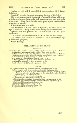
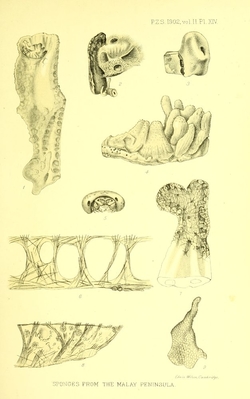
![Surface covered with low coiiuli 1-2 mm. apart and 0’2-0’4mm. in height. Oscula (?) minute, inconspicuous near the tips of the lobes. The skeleton consists of a network of very thin fibres, which are not distinguishable into main and secondary, and are uniformly areniferous. They form for the most part square meshes about 0*64 mm. in breadth. Fibres 0*03-0'06 mm. thick. The ectosome is a thin layer of cystenchyma, thickest at tlie tips of the lobes. Only in this layer are spongoblasts obvious. Spermatozoa are present in vaiious stages and in great abundance. The ciliated chambers measure *07 x '04 mm. on an average. The whole choanosome is peiineated by a filamentous alga (? Oscillaria spongeMa^). Great Redang. EXPLANATION OF THE PLATES. Plate XIV. Fig. 1. One of the digitiform processes of Ciocali/pta melichlora, p. 214. Nat. size. 2. Cinachyra malaccensis (p. 219), slightly larger than nat. size. 3. Spirastrella inconstans (p. 216), attacheci to a fragment of stone. Nat. size. 4. Spongelia digitata (p. 220), attached to a liranch of dead coral. Nat. size. 5. Heniera sp. 1 (p. 210), growing on a crab. X 2. 6. A section oi Pseudosuherites cava (p. 217). X 15. 7. Ciocalypta rutila (p. 215), almost the entire specimen. X *. 8. Psperella sulevoidea (p. 213), in section. X 20. 9. P. sulevoidea (p. 213), entire specimen. X f. Plate XV. Fig. 1. Microcalthrops and spined microxea of Dereitus pauper, p. 218. 2. One complete mesh of the skeleton of Spongelia digitata, p. 220. 3. Meniera sp. 5 (p. 211). a, node of the skeletal network; h, egg; c, ciliated chamber; d, isolated spicule; e, piece of fibre with chaplet-cells ; f, isolated chaplet-cell; f, its segment of fibre. 4. Spicules of Suberites laxosuherites, p. 217. 6. Megascleres of Cinachyra malaccensis, p. 219. 6. Sigmata and centrangulate sigmata of Gellius centrangulatus, p. 212. 7. Sigmata, toxa, and centrangulate sigmata of Gellius Sagittarius, p. 212. 8. Spicules of Ciocalypta melichlora, p. 214. 9. Spicules of Biemma democratica, p. 213. 10. Spicules of JSsperella sulevoidea, p. 213. 11. Spicules of Eeniera sp. 2, p. 210. [13]](https://iiif.wellcomecollection.org/image/b22406414_0017.jp2/full/800%2C/0/default.jpg)