On the sponges collected during the 'Skeat expedition' to the Malay peninsula, 1899-1900 / by Igerna B.J. Sollas.
- Sollas, Igerna Brünhilda Johnson.
- Date:
- 1902
Licence: In copyright
Credit: On the sponges collected during the 'Skeat expedition' to the Malay peninsula, 1899-1900 / by Igerna B.J. Sollas. Source: Wellcome Collection.
Provider: This material has been provided by The Royal College of Surgeons of England. The original may be consulted at The Royal College of Surgeons of England.
4/20 (page 210)
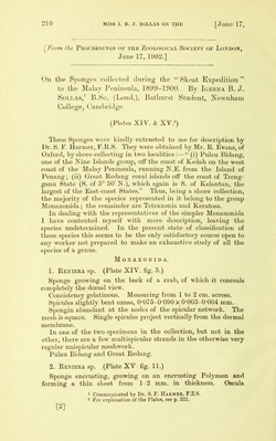
![^From the PiiocBifidings or the Zoological Society or London, June 17, 1902.] On the S])onges collected during the “ Skeat Expedition” to the Malay Peninsula, 1899-1900. By Igekna B. J. SoLLAS,^ B.Sc. (Loud.), Bathurst Student, Newnhani College, Cainhridge. (Plates XIV. & XV.^) These Sponges wei e kindly entrusted to me for description by Dr. S. F. Hariner, F.ll.S. They were obtained by Mr. R. Evans, of Oxford, by shore-collecting in two localities :—“ (i) Pulau Bidang, one of the Nine Islands group, otf the coast of Kedah on the west coast of the Malay Peninsula, running N.E. from the Island of Penang; (ii) Great Reclang coral islands ofi' the coast of Tieng- ganu State (S. of 5° 50' N.), which again is S. of Kelantan, the largest of the East-coast States.” Thus, being a shore collection, the majoiity of the species represented in it belong to the group IMonaxonida ; the remainder ai-e Tetraxonia and Kei*atosa. In dealing with the representatives of the simpler Monaxonida I have contented myself with mere description, leaving the species undeteiinined. In the piesent state of classification of these species this seems to be the only satisfactoiy course open to any worker not prepared to make an exhaustive study of all the species of a genus. Monaxonida. 1. Reniera sp. (Plate XIV. fig. 5.) Sponge growing on the back of a crab, of which it conceals completely the dorsal view. Consistency gelatinous. Measuring from 1 to 2 cm. across. Spicules slightly bent oxeas, 0*075-0*090x 0’003-0*004 mm. Spongin abundant at the nodes of the spicular network. The mesh is square. Single spicules project vertically from the dermal membrane. In one of the two specimens in the collection, but not in the other, there are a few multispicular strands in the otherwise very regular unispicular meshwork. Pulau Bidang and Great Redang. 2. Reniera sp. (Plate XV-fig. 11.) Sponge encrusting, growing on an encrusting Polyzoon and forming a thin sheet from 1-2 mm. in thickness. Oscula ^ Commuiiicated by Dr. S. F. Haemee, F.Z.S. 2 For explanation of the Plates, see p. 221. [2]](https://iiif.wellcomecollection.org/image/b22406414_0006.jp2/full/800%2C/0/default.jpg)